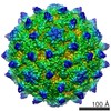
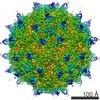
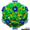
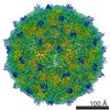
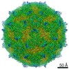
ジャーナル: J Virol / 年: 2014 タイトル: Kinetic and structural analysis of coxsackievirus B3 receptor interactions and formation of the A-particle. 著者: Lindsey J Organtini / Alexander M Makhov / James F Conway / Susan Hafenstein / Steven D Carson / 要旨: The coxsackievirus and adenovirus receptor (CAR) has been identified as the cellular receptor for group B coxsackieviruses, including serotype 3 (CVB3). CAR mediates infection by binding to CVB3 and ...The coxsackievirus and adenovirus receptor (CAR) has been identified as the cellular receptor for group B coxsackieviruses, including serotype 3 (CVB3). CAR mediates infection by binding to CVB3 and catalyzing conformational changes in the virus that result in formation of the altered, noninfectious A-particle. Kinetic analyses show that the apparent first-order rate constant for the inactivation of CVB3 by soluble CAR (sCAR) at physiological temperatures varies nonlinearly with sCAR concentration. Cryo-electron microscopy (cryo-EM) reconstruction of the CVB3-CAR complex resulted in a 9.0-Å resolution map that was interpreted with the four available crystal structures of CAR, providing a consensus footprint for the receptor binding site. The analysis of the cryo-EM structure identifies important virus-receptor interactions that are conserved across picornavirus species. These conserved interactions map to variable antigenic sites or structurally conserved regions, suggesting a combination of evolutionary mechanisms for receptor site preservation. The CAR-catalyzed A-particle structure was solved to a 6.6-Å resolution and shows significant rearrangement of internal features and symmetric interactions with the RNA genome. IMPORTANCE: This report presents new information about receptor use by picornaviruses and highlights the importance of attaining at least an ∼9-Å resolution for the interpretation of cryo-EM ...IMPORTANCE: This report presents new information about receptor use by picornaviruses and highlights the importance of attaining at least an ∼9-Å resolution for the interpretation of cryo-EM complex maps. The analysis of receptor binding elucidates two complementary mechanisms for preservation of the low-affinity (initial) interaction of the receptor and defines the kinetics of receptor-catalyzed conformational change to the A-particle.
名称: Coxsackievirus B3 A-Particle / タイプ: sample / ID: 1000 / 詳細: purified virus and receptor complex in solution / 集合状態: icosahedral virus / Number unique components: 1
分子量
実験値: 7 MDa
-
超分子 #1: Human coxsackievirus B3
超分子
名称: Human coxsackievirus B3 / タイプ: virus / ID: 1 / Name.synonym: CVB3 詳細: Virus was incubated with excess CAR at 4 degrees C then transitioned to 37 degrees C in order to create the A-particle. NCBI-ID: 12072 / 生物種: Human coxsackievirus B3 / Sci species strain: CVB3/28 / データベース: NCBI / ウイルスタイプ: VIRION / ウイルス・単離状態: STRAIN / ウイルス・エンベロープ: No / ウイルス・中空状態: No / Syn species name: CVB3
 ムービー
ムービー コントローラー


 データを開く
 基本情報
基本情報 マップデータ
 試料
試料 キーワード
キーワード
 Human coxsackievirus B3 (コクサッキーウイルス)
 データ登録者
 引用
引用
 構造の表示
構造の表示 ムービービューア
 ダウンロードとリンク
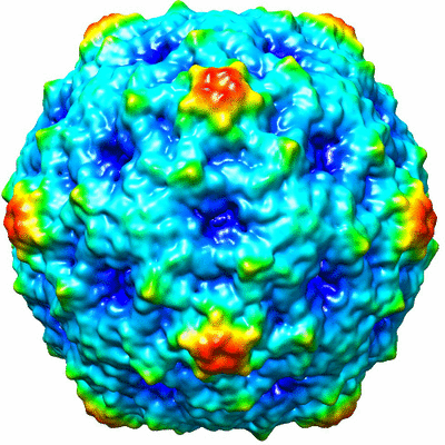 400_5928.gif
 80_5928.gif
 http://ftp.pdbj.org/pub/emdb/structures/EMD-5928
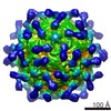

 Z (Sec.)
 Y (Row.)
 X (Col.)


 試料の構成要素
 Homo sapiens (ヒト) / 別称: VERTEBRATES
 解析
解析 電子顕微鏡法
 FIELD EMISSION GUN
