ムービー
ムービー コントローラー


+ データを開く

- 基本情報
基本情報
| 登録情報 | データベース: EMDB / ID: EMD-1710 | |||||||||
|---|---|---|---|---|---|---|---|---|---|---|
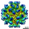
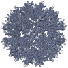
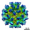
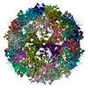
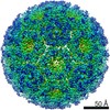
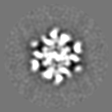
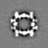
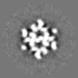
| タイトル | Cryo-EM 3D model of the icosahedral particle composed of Rous sarcoma virus capsid protein pentamers | |||||||||
 マップデータ マップデータ | This is a 3D reconstruction of the icosahedral particle composed of Rous sarcoma virus capsid protein pentamers | |||||||||
 試料 試料 |
| |||||||||
| 機能・相同性 |  機能・相同性情報 機能・相同性情報host cell nucleoplasm / viral procapsid maturation / host cell nucleolus / 加水分解酵素; プロテアーゼ; ペプチド結合加水分解酵素; アスパラギン酸プロテアーゼ / viral capsid / structural constituent of virion / aspartic-type endopeptidase activity / nucleic acid binding / viral translational frameshifting / host cell plasma membrane ...host cell nucleoplasm / viral procapsid maturation / host cell nucleolus / 加水分解酵素; プロテアーゼ; ペプチド結合加水分解酵素; アスパラギン酸プロテアーゼ / viral capsid / structural constituent of virion / aspartic-type endopeptidase activity / nucleic acid binding / viral translational frameshifting / host cell plasma membrane / proteolysis / zinc ion binding / membrane 類似検索 - 分子機能 | |||||||||
| 生物種 |  Rous sarcoma virus - Prague C (ラウス肉腫ウイルス) Rous sarcoma virus - Prague C (ラウス肉腫ウイルス) | |||||||||
| 手法 | 単粒子再構成法 / クライオ電子顕微鏡法 / 解像度: 18.3 Å | |||||||||
 データ登録者 データ登録者 | Hyun JK / Radjainia M / Kingston RL / Mitra AK | |||||||||
 引用 引用 | ジャーナル: J Biol Chem / 年: 2010 タイトル: Proton-driven assembly of the Rous Sarcoma virus capsid protein results in the formation of icosahedral particles. 著者: Jae-Kyung Hyun / Mazdak Radjainia / Richard L Kingston / Alok K Mitra /  要旨: In a mature and infectious retroviral particle, the capsid protein (CA) forms a shell surrounding the genomic RNA and the replicative machinery of the virus. The irregular nature of this capsid shell ...In a mature and infectious retroviral particle, the capsid protein (CA) forms a shell surrounding the genomic RNA and the replicative machinery of the virus. The irregular nature of this capsid shell precludes direct atomic resolution structural analysis. CA hexamers and pentamers are the fundamental building blocks of the capsid, however the pentameric state, in particular, remains poorly characterized. We have developed an efficient in vitro protocol for studying the assembly of Rous sarcoma virus (RSV) CA that involves mild acidification and produces structures modeling the authentic viral capsid. These structures include regular spherical particles with T = 1 icosahedral symmetry, built from CA pentamers alone. These particles were subject to cryoelectron microscopy (cryo-EM) and image processing, and a pseudo-atomic model of the icosahedron was created by docking atomic structures of the constituent CA domains into the cryo-EM-derived three-dimensional density map. The N-terminal domain (NTD) of CA forms pentameric turrets, which decorate the surface of the icosahedron, while the C-terminal domain (CTD) of CA is positioned underneath, linking the pentamers. Biophysical analysis of the icosahedral particle preparation reveals that CA monomers and icosahedra are the only detectable species and that these exist in reversible equilibrium at pH 5. These same acidic conditions are known to promote formation of a RSV CA CTD dimer, present within the icosahedral particle, which facilitates capsid assembly. The results are consistent with a model in which RSV CA assembly is a nucleation-limited process driven by very weak protein-protein interactions. | |||||||||
| 履歴 |
|
- 構造の表示
構造の表示
| ムービー |
ムービービューア |
|---|---|
| 構造ビューア | EMマップ: SurfViewMolmilJmol/JSmol |
| 添付画像 |
 UCSF Chimera
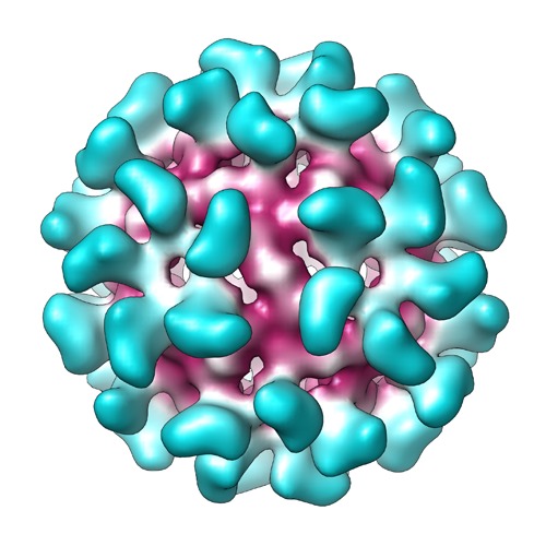
- ダウンロードとリンク
-EMDBアーカイブ
| マップデータ | emd_1710.map.gz | 7.4 MB | EMDBマップデータ形式 | |
|---|---|---|---|---|
| ヘッダ (付随情報) | emd-1710-v30.xmlemd-1710.xml | 10.7 KB 10.7 KB | 表示 表示 | EMDBヘッダ |
| 画像 | map_EMD-1710.tif | 758.4 KB | ||
| アーカイブディレクトリ |  http://ftp.pdbj.org/pub/emdb/structures/EMD-1710ftp://ftp.pdbj.org/pub/emdb/structures/EMD-1710 http://ftp.pdbj.org/pub/emdb/structures/EMD-1710ftp://ftp.pdbj.org/pub/emdb/structures/EMD-1710 | HTTPS FTP |
-検証レポート
| 文書・要旨 | emd_1710_validation.pdf.gz | 236.8 KB | 表示 | EMDB検証レポート |
|---|---|---|---|---|
| 文書・詳細版 | emd_1710_full_validation.pdf.gz | 235.9 KB | 表示 | |
| XML形式データ | emd_1710_validation.xml.gz | 5.6 KB | 表示 | |
| アーカイブディレクトリ | https://ftp.pdbj.org/pub/emdb/validation_reports/EMD-1710ftp://ftp.pdbj.org/pub/emdb/validation_reports/EMD-1710 | HTTPS FTP |
-関連構造データ

-リンク
| EMDBのページ | EMDB (EBI/PDBe) / EMDataResource |
|---|
-マップ
| ファイル | ダウンロード / ファイル: emd_1710.map.gz / 形式: CCP4 / 大きさ: 15.3 MB / タイプ: IMAGE STORED AS FLOATING POINT NUMBER (4 BYTES) | ||||||||||||||||||||||||||||||||||||||||||||||||||||||||||||||||||||
|---|---|---|---|---|---|---|---|---|---|---|---|---|---|---|---|---|---|---|---|---|---|---|---|---|---|---|---|---|---|---|---|---|---|---|---|---|---|---|---|---|---|---|---|---|---|---|---|---|---|---|---|---|---|---|---|---|---|---|---|---|---|---|---|---|---|---|---|---|---|
| 注釈 | This is a 3D reconstruction of the icosahedral particle composed of Rous sarcoma virus capsid protein pentamers | ||||||||||||||||||||||||||||||||||||||||||||||||||||||||||||||||||||
| 投影像・断面図 | 画像のコントロール
画像は Spider により作成 | ||||||||||||||||||||||||||||||||||||||||||||||||||||||||||||||||||||
| ボクセルのサイズ | X=Y=Z: 2.5 Å | ||||||||||||||||||||||||||||||||||||||||||||||||||||||||||||||||||||
| 密度 |
| ||||||||||||||||||||||||||||||||||||||||||||||||||||||||||||||||||||
| 対称性 | 空間群: 1 | ||||||||||||||||||||||||||||||||||||||||||||||||||||||||||||||||||||
| 詳細 | EMDB XML:
CCP4マップ ヘッダ情報:
| ||||||||||||||||||||||||||||||||||||||||||||||||||||||||||||||||||||
 Z (Sec.)
 Y (Row.)
 X (Col.)


-添付データ
- 試料の構成要素
-全体 : Icosahedral particles composed of Rous sarcoma virus capsid protein
| 全体 | 名称: Icosahedral particles composed of Rous sarcoma virus capsid protein |
|---|---|
| 要素 |
|
-超分子 #1000: Icosahedral particles composed of Rous sarcoma virus capsid protein
| 超分子 | 名称: Icosahedral particles composed of Rous sarcoma virus capsid protein タイプ: sample / ID: 1000 詳細: The icosahedral particles were assembled in vitro, by transferring recombinant Rous sarcoma virus capsid protein monomers into high salt, mildly acidic buffer 集合状態: Icosahedral particle containing 12 CA pentamers (i.e. 60 monomers) Number unique components: 1 |
|---|---|
| 分子量 | 理論値: 1.5 MDa |
-分子 #1: Capsid protein p27
| 分子 | 名称: Capsid protein p27 / タイプ: protein_or_peptide / ID: 1 / Name.synonym: Capsid protein p27 / コピー数: 60 集合状態: Icosahedral particle composed of 12 protein pentamers 組換発現: Yes |
|---|---|
| 由来(天然) | 生物種: Rous sarcoma virus - Prague C (ラウス肉腫ウイルス) |
| 分子量 | 理論値: 1.53 MDa |
| 組換発現 | 生物種:  |
-実験情報
-構造解析
| 手法 | クライオ電子顕微鏡法 |
|---|---|
 解析 解析 | 単粒子再構成法 |
| 試料の集合状態 | particle |
-試料調製
| 濃度 | 1.2 mg/mL |
|---|---|
| 緩衝液 | pH: 5 詳細: 0.1M citric acid, 5mM MOPS/KOH, 725mM NaCl, 0.25mM Na azide, 0.125mM TCEP-HCl |
| グリッド | 詳細: Holey carbon 400 mesh copper grid |
| 凍結 | 凍結剤: ETHANE / チャンバー内湿度: 90 % / チャンバー内温度: 85 K / 装置: FEI VITROBOT MARK IV / 詳細: Vitrification instrument: Vitrobot Mark IV / 手法: Blot for 5 seconds before plunging |
- 電子顕微鏡法
| 顕微鏡 | FEI TECNAI 12 |
|---|---|
| 温度 | 平均: 103 K |
| アライメント法 | Legacy - 非点収差: Objective lens astigmatism was corrected at 60,000 - 140,000 times magnification using live fft |
| 撮影 | カテゴリ: FILM / フィルム・検出器のモデル: KODAK SO-163 FILM デジタル化 - スキャナー: NIKON SUPER COOLSCAN 9000 デジタル化 - サンプリング間隔: 10.5 µm / 実像数: 21 / 平均電子線量: 18 e/Å2 / ビット/ピクセル: 8 |
| 電子線 | 加速電圧: 120 kV / 電子線源: LAB6 |
| 電子光学系 | 照射モード: FLOOD BEAM / 撮影モード: BRIGHT FIELD / Cs: 2.0 mm / 最大 デフォーカス(公称値): 3.0 µm / 最小 デフォーカス(公称値): 0.8 µm / 倍率(公称値): 42000 |
| 試料ステージ | 試料ホルダー: Side entry liquid nitrogen-cooled cryo specimen holder 試料ホルダーモデル: GATAN LIQUID NITROGEN |
-画像解析
| CTF補正 | 詳細: Each micrograph |
|---|---|
| 最終 再構成 | 想定した対称性 - 点群: I (正20面体型対称) / アルゴリズム: OTHER / 解像度のタイプ: BY AUTHOR / 解像度: 18.3 Å / 解像度の算出法: FSC 0.5 CUT-OFF / ソフトウェア - 名称: Bsoft, PFT2, EM3DR2 詳細: The digitized micrographs were processed using Bsoft. Orientation and origin search of the particles and 3D reconstruction were performed using PFT2 and EM3DR, respectively 使用した粒子像数: 1310 |
-原子モデル構築 1
| 初期モデル | (PDB ID: , ) |
|---|---|
| ソフトウェア | 名称: Chimera,Sculptor |
| 詳細 | Protocol: Rigid body. The domain structures were manually fitted into the 3D reconstruction, and then the fitting was refined using Sculptor |
| 精密化 | 空間: RECIPROCAL / プロトコル: RIGID BODY FIT / 当てはまり具合の基準: Cross-correlation |
| 得られたモデル |  PDB-2x8q: |