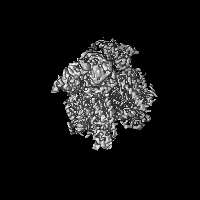
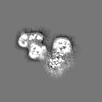
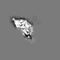
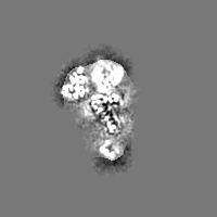
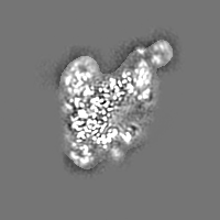
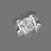
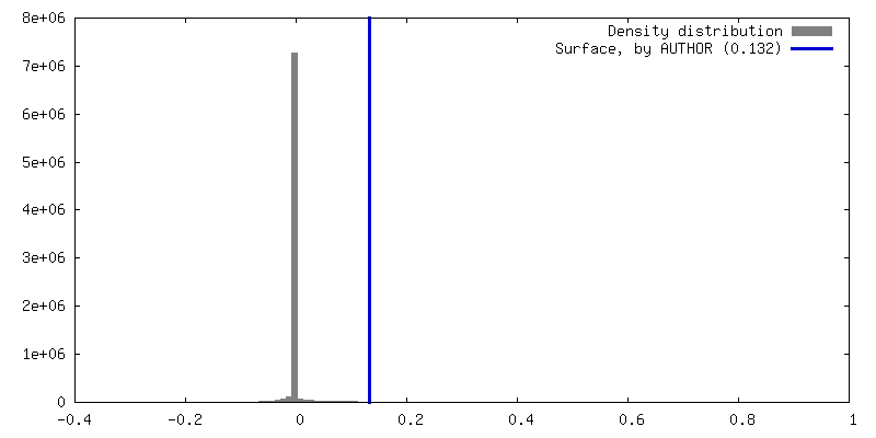
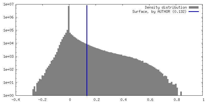
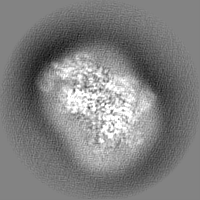
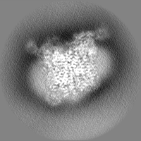
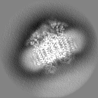
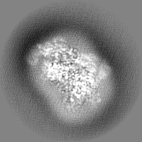
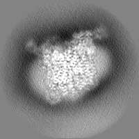
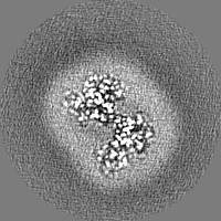
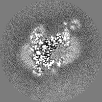
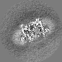
- EMDB-14224: 2.8 Angstrom cryo-EM structure of the dimeric cytochrome b6f-PetP... -
+
データを開く
IDまたはキーワード:
読み込み中...
-
基本情報
登録情報
データベース: EMDB / ID: EMD-14224
タイトル
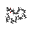
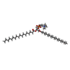
2.8 Angstrom cryo-EM structure of the dimeric cytochrome b6f-PetP complex from Synechocystis sp. PCC 6803 with natively bound lipids and plastoquinone molecules
マップデータ
試料
複合体: Cytochrome b6f complex in association with PetPシトクロムb6f複合体
タンパク質・ペプチド: x 9種
リガンド: x 13種
機能・相同性
機能・相同性情報
: / シトクロムb6f複合体 / electron transporter, transferring electrons within cytochrome b6/f complex of photosystem II activity / cytochrome complex assembly / photosynthetic electron transport chain / plasma membrane-derived thylakoid membrane / electron transporter, transferring electrons within the cyclic electron transport pathway of photosynthesis activity / 光合成 / respiratory electron transport chain / electron transfer activity ...: / シトクロムb6f複合体 / electron transporter, transferring electrons within cytochrome b6/f complex of photosystem II activity / cytochrome complex assembly / photosynthetic electron transport chain / plasma membrane-derived thylakoid membrane / electron transporter, transferring electrons within the cyclic electron transport pathway of photosynthesis activity / 光合成 / respiratory electron transport chain / electron transfer activity / membrane => GO:0016020 / iron ion binding / heme binding / metal ion binding 類似検索 - 分子機能
Biotechnology and Biological Sciences Research Council (BBSRC)
BB/M011151/1
英国
Biotechnology and Biological Sciences Research Council (BBSRC)
BB/V006630/1
英国
Leverhulme Trust
RPG-2019-045
英国
Wellcome Trust
nr21005
英国
引用
ジャーナル: Biochem J / 年: 2022 タイトル: Cryo-EM structures of the Synechocystis sp. PCC 6803 cytochrome b6f complex with and without the regulatory PetP subunit. 著者: Matthew S Proctor / Lorna A Malone / David A Farmer / David J K Swainsbury / Frederick R Hawkings / Federica Pastorelli / Thomas Z Emrich-Mills / C Alistair Siebert / C Neil Hunter / Matthew ...著者: Matthew S Proctor / Lorna A Malone / David A Farmer / David J K Swainsbury / Frederick R Hawkings / Federica Pastorelli / Thomas Z Emrich-Mills / C Alistair Siebert / C Neil Hunter / Matthew P Johnson / Andrew Hitchcock / 要旨: In oxygenic photosynthesis, the cytochrome b6f (cytb6f) complex links the linear electron transfer (LET) reactions occurring at photosystems I and II and generates a transmembrane proton gradient via ...In oxygenic photosynthesis, the cytochrome b6f (cytb6f) complex links the linear electron transfer (LET) reactions occurring at photosystems I and II and generates a transmembrane proton gradient via the Q-cycle. In addition to this central role in LET, cytb6f also participates in a range of processes including cyclic electron transfer (CET), state transitions and photosynthetic control. Many of the regulatory roles of cytb6f are facilitated by auxiliary proteins that differ depending upon the species, yet because of their weak and transient nature the structural details of these interactions remain unknown. An apparent key player in the regulatory balance between LET and CET in cyanobacteria is PetP, a ∼10 kDa protein that is also found in red algae but not in green algae and plants. Here, we used cryogenic electron microscopy to determine the structure of the Synechocystis sp. PCC 6803 cytb6f complex in the presence and absence of PetP. Our structures show that PetP interacts with the cytoplasmic side of cytb6f, displacing the C-terminus of the PetG subunit and shielding the C-terminus of cytochrome b6, which binds the heme cn cofactor that is suggested to mediate CET. The structures also highlight key differences in the mode of plastoquinone binding between cyanobacterial and plant cytb6f complexes, which we suggest may reflect the unique combination of photosynthetic and respiratory electron transfer in cyanobacterial thylakoid membranes. The structure of cytb6f from a model cyanobacterial species amenable to genetic engineering will enhance future site-directed mutagenesis studies of structure-function relationships in this crucial ET complex.
超分子 #1: Cytochrome b6f complex in association with PetP
超分子
名称: Cytochrome b6f complex in association with PetP / タイプ: complex / キメラ: Yes / ID: 1 / 親要素: 0 / 含まれる分子: #1-#9 詳細: strep-PetA used to purify cytochrome b6f complex from Synechocystis sp. PCC 6803. 6xHis-PetP recombinantly expressed in E. coli used to pull down cytochrome b6f complex and form the cytochrome b6f-PetP complex.
由来(天然)
生物種: Synechocystis sp. PCC 6803 (バクテリア)
+
分子 #1: Uncharacterized protein Cp097, conserved in cyanobacteria
分子
名称: Uncharacterized protein Cp097, conserved in cyanobacteria タイプ: protein_or_peptide / ID: 1 / コピー数: 2 / 光学異性体: LEVO
PDB-7r0w: 2.8 Angstrom cryo-EM structure of the dimeric cytochrome b6f-PetP complex from Synechocystis sp. PCC 6803 with natively bound lipids and plastoquinone molecules
 ムービー
ムービー コントローラー


 データを開く
 基本情報
基本情報
 マップデータ
 試料
試料 機能・相同性情報
 : /
: / 
 データ登録者
 英国, 4件
  引用
引用 構造の表示
構造の表示
 ダウンロードとリンク
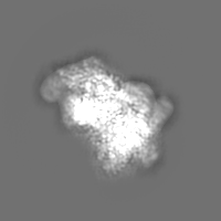
 emd_14224.png
 http://ftp.pdbj.org/pub/emdb/structures/EMD-14224


 Z (Sec.)
 Y (Row.)
 X (Col.)


 試料の構成要素


 解析
解析 電子顕微鏡法