 ムービー
ムービー コントローラー


+ データを開く

- 基本情報
基本情報
| 登録情報 | データベース: EMDB / ID: EMD-1422 | |||||||||
|---|---|---|---|---|---|---|---|---|---|---|
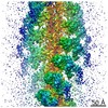
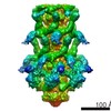
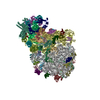
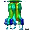
| タイトル | Structure and composition of the Shigella flexneri "needle complex", a part of its type III secreton. | |||||||||
 マップデータ マップデータ | Basal Body of Shigella flexneri T3SS - needle removed | |||||||||
 試料 試料 |
| |||||||||
| 機能・相同性 | : / protein binding / Type III secretion system outer membrane pore YscC/HrcC / Type III secretion system, needle protein / Flagellar M-ring , N-terminal 機能・相同性情報 機能・相同性情報 | |||||||||
| 生物種 |  Shigella flexneri (フレクスナー赤痢菌) Shigella flexneri (フレクスナー赤痢菌) | |||||||||
| 手法 | 単粒子再構成法 / クライオ電子顕微鏡法 / ネガティブ染色法 / 解像度: 17.0 Å | |||||||||
 データ登録者 データ登録者 | Blocker AJ / Jouihri N / Larquet E / Gounon P / Ebel F / Parsot C / Sansonetti P / Allaoui A | |||||||||
 引用 引用 | ジャーナル: Mol Microbiol / 年: 2001 タイトル: Structure and composition of the Shigella flexneri "needle complex", a part of its type III secreton. 著者: A Blocker / N Jouihri / E Larquet / P Gounon / F Ebel / C Parsot / P Sansonetti / A Allaoui /  要旨: Type III secretion systems (TTSSs or secretons), essential virulence determinants of many Gram-negative bacteria, serve to translocate proteins directly from the bacteria into the host cytoplasm. ...Type III secretion systems (TTSSs or secretons), essential virulence determinants of many Gram-negative bacteria, serve to translocate proteins directly from the bacteria into the host cytoplasm. Electron microscopy (EM) indicates that the TTSSs of Shigella flexneri are composed of: (1) an external needle; (2) a transmembrane domain; and (3) a cytoplasmic bulb. EM analysis of purified and negatively stained parts 1, 2 and a portion of 3 of the TTSS, together termed the "needle complex" (NC), produced an average image at 17 A resolution in which a base, an outer ring and a needle, inserted through the ring into the base, could be discerned. This analysis and cryoEM images of NCs indicated that the needle and base contain a central 2-3 nm canal. Five major NC components, MxiD, MxiG, MxiJ, MxiH and MxiI, were identified by N-terminal sequencing. MxiG and MxiJ are predicted to be inner membrane proteins and presumably form the base. MxiD is predicted to be an outer membrane protein and to form the outer ring. MxiH and MxiI are small hydrophilic proteins. Mutants lacking either of these proteins formed needleless secretons and were unable to secrete Ipa proteins. As MxiH was present in NCs in large molar excess, we propose that it is the major needle component. MxiI may cap at the external needle tip. | |||||||||
| 履歴 |
|
- 構造の表示
構造の表示
| ムービー |
ムービービューア |
|---|---|
| 構造ビューア | EMマップ: SurfViewMolmilJmol/JSmol |
| 添付画像 |
 UCSF Chimera
- ダウンロードとリンク
-EMDBアーカイブ
| マップデータ | emd_1422.map.gz | 3.3 MB | EMDBマップデータ形式 | |
|---|---|---|---|---|
| ヘッダ (付随情報) | emd-1422-v30.xmlemd-1422.xml | 14.9 KB 14.9 KB | 表示 表示 | EMDBヘッダ |
| 画像 |  1422.gif 1422.gif | 17.5 KB | ||
| アーカイブディレクトリ |  http://ftp.pdbj.org/pub/emdb/structures/EMD-1422ftp://ftp.pdbj.org/pub/emdb/structures/EMD-1422 http://ftp.pdbj.org/pub/emdb/structures/EMD-1422ftp://ftp.pdbj.org/pub/emdb/structures/EMD-1422 | HTTPS FTP |
-検証レポート
| 文書・要旨 | emd_1422_validation.pdf.gz | 199.4 KB | 表示 | EMDB検証レポート |
|---|---|---|---|---|
| 文書・詳細版 | emd_1422_full_validation.pdf.gz | 198.5 KB | 表示 | |
| XML形式データ | emd_1422_validation.xml.gz | 7 KB | 表示 | |
| アーカイブディレクトリ | https://ftp.pdbj.org/pub/emdb/validation_reports/EMD-1422ftp://ftp.pdbj.org/pub/emdb/validation_reports/EMD-1422 | HTTPS FTP |
-関連構造データ
-リンク
| EMDBのページ | EMDB (EBI/PDBe) / EMDataResource |
|---|
-マップ
| ファイル | ダウンロード / ファイル: emd_1422.map.gz / 形式: CCP4 / 大きさ: 100.6 MB / タイプ: IMAGE STORED AS FLOATING POINT NUMBER (4 BYTES) | ||||||||||||||||||||||||||||||||||||||||||||||||||||||||||||||||||||
|---|---|---|---|---|---|---|---|---|---|---|---|---|---|---|---|---|---|---|---|---|---|---|---|---|---|---|---|---|---|---|---|---|---|---|---|---|---|---|---|---|---|---|---|---|---|---|---|---|---|---|---|---|---|---|---|---|---|---|---|---|---|---|---|---|---|---|---|---|---|
| 注釈 | Basal Body of Shigella flexneri T3SS - needle removed | ||||||||||||||||||||||||||||||||||||||||||||||||||||||||||||||||||||
| ボクセルのサイズ | X=Y=Z: 2.2 Å | ||||||||||||||||||||||||||||||||||||||||||||||||||||||||||||||||||||
| 密度 |
| ||||||||||||||||||||||||||||||||||||||||||||||||||||||||||||||||||||
| 対称性 | 空間群: 1 | ||||||||||||||||||||||||||||||||||||||||||||||||||||||||||||||||||||
| 詳細 | EMDB XML:
CCP4マップ ヘッダ情報:
| ||||||||||||||||||||||||||||||||||||||||||||||||||||||||||||||||||||
-添付データ
- 試料の構成要素
-全体 : needle complex or basal body of the Shigella flexneri T3SS
| 全体 | 名称: needle complex or basal body of the Shigella flexneri T3SS |
|---|---|
| 要素 |
|
-超分子 #1000: needle complex or basal body of the Shigella flexneri T3SS
| 超分子 | 名称: needle complex or basal body of the Shigella flexneri T3SS タイプ: sample / ID: 1000 / 集合状態: not yet fully determined / Number unique components: 5 |
|---|
-分子 #1: MxiH
| 分子 | 名称: MxiH / タイプ: protein_or_peptide / ID: 1 / 詳細: none; copy number is approximative / コピー数: 120 / 集合状態: helical polymer / 組換発現: No / データベース: NCBI |
|---|---|
| 由来(天然) | 生物種: Shigella flexneri (フレクスナー赤痢菌) / 株: M90T / 組織: bacterium / Organelle: T3SS / 細胞中の位置: extracellular |
| 分子量 | 実験値: 9.265 MDa / 理論値: 9.265 MDa |
| 配列 | InterPro: Type III secretion system, needle protein |
-分子 #2: MxiI
| 分子 | 名称: MxiI / タイプ: protein_or_peptide / ID: 2 / 詳細: none; Copy number is approximative / コピー数: 20 / 集合状態: probably helical / 組換発現: No / データベース: NCBI |
|---|---|
| 由来(天然) | 生物種: Shigella flexneri (フレクスナー赤痢菌) / 株: M90T / 細胞: bacterium / Organelle: T3SS / 細胞中の位置: periplasmic |
| 分子量 | 実験値: 10.633 MDa / 理論値: 10.633 MDa |
-分子 #3: MxiD
| 分子 | 名称: MxiD / タイプ: protein_or_peptide / ID: 3 詳細: experimental weight is theoretical weight, with predicted signal sequence removed; oligomeric state currently unknown 集合状態: oligomer / 組換発現: No / データベース: NCBI |
|---|---|
| 由来(天然) | 生物種: Shigella flexneri (フレクスナー赤痢菌) / 株: M90T / 細胞: bacterium / Organelle: T3SS / 細胞中の位置: outer membrane |
| 分子量 | 実験値: 63.218 MDa / 理論値: 60.749 MDa |
| 配列 | GO: GO: 0015448 InterPro: Type III secretion system outer membrane pore YscC/HrcC |
-分子 #4: MxiG
| 分子 | 名称: MxiG / タイプ: protein_or_peptide / ID: 4 詳細: no signal sequence cleavage in this protein; oligomeric state unknown 集合状態: oligomer / 組換発現: No / データベース: NCBI |
|---|---|
| 由来(天然) | 生物種: Shigella flexneri (フレクスナー赤痢菌) / 株: M90T / 細胞: bacterium / Organelle: T3SS / 細胞中の位置: inner membrane |
| 分子量 | 実験値: 43.002 MDa / 理論値: 43.002 MDa |
| 配列 | GO: protein binding |
-分子 #5: MxiJ
| 分子 | 名称: MxiJ / タイプ: protein_or_peptide / ID: 5 詳細: experimental weight is theoretical weight, with predicted signal sequence removed; oligomeric state unknown 集合状態: oligomer / 組換発現: No / データベース: NCBI |
|---|---|
| 由来(天然) | 生物種: Shigella flexneri (フレクスナー赤痢菌) / 株: M90T / 細胞: bacterium / Organelle: T3SS / 細胞中の位置: inner membrane |
| 分子量 | 実験値: 27.509 MDa / 理論値: 25.488 MDa |
| 配列 | GO: protein binding / InterPro: Flagellar M-ring , N-terminal |
-実験情報
-構造解析
| 手法 | ネガティブ染色法, クライオ電子顕微鏡法 |
|---|---|
 解析 解析 | 単粒子再構成法 |
| 試料の集合状態 | particle |
-試料調製
| 緩衝液 | pH: 7.4 詳細: samples made in 50 mM Tris pH 8, 5 mM EDTA, 0.1 % v/v Triton X-100 and diluted 1:5 in 20 mM Tris pH 7.4 |
|---|---|
| 染色 | タイプ: NEGATIVE / 詳細: 1% uranyl acetate pH 7.5 |
| グリッド | 詳細: carbon-coated glow-discharged copper grids |
| 凍結 | 凍結剤: ETHANE |
- 電子顕微鏡法
| 顕微鏡 | FEI/PHILIPS CM120T |
|---|---|
| 温度 | 最低: 293 K / 最高: 293 K / 平均: 293 K |
| 詳細 | low-dose mode used, imaging done in 1999-2000 |
| 撮影 | カテゴリ: FILM / フィルム・検出器のモデル: KODAK SO-163 FILM / デジタル化 - スキャナー: OTHER / デジタル化 - サンプリング間隔: 10 µm / 実像数: 20 / 平均電子線量: 10 e/Å2 詳細: Hi-Scan rotary drum microdensitometer used, final resolution was 2.2 A/pixel (not 5 A/ pixel as incorrectly stated in Materials and Methods of paper) |
| 電子線 | 加速電圧: 100 kV / 電子線源: LAB6 |
| 電子光学系 | 照射モード: SPOT SCAN / 撮影モード: BRIGHT FIELD / Cs: 2 mm / 最大 デフォーカス(公称値): 1.2 µm / 最小 デフォーカス(公称値): 0.6 µm / 倍率(公称値): 45000 |
| 試料ステージ | 試料ホルダー: Eucentric / 試料ホルダーモデル: OTHER |
-画像解析
| 詳細 | Particles selected using interactive WEB selection program |
|---|---|
| 最終 再構成 | アルゴリズム: OTHER / 解像度のタイプ: BY AUTHOR / 解像度: 17.0 Å / 解像度の算出法: OTHER / ソフトウェア - 名称: SPIDER 詳細: 1) 2D-average>iterative multireference alignment process (Boekema et al, 1986), using Ward's merging criterion until stable classes obtained; 2) 3D reconstruction> cylindrical symmetry was ...詳細: 1) 2D-average>iterative multireference alignment process (Boekema et al, 1986), using Ward's merging criterion until stable classes obtained; 2) 3D reconstruction> cylindrical symmetry was assumed and the final two dimensional projection converted to a three-dimensional map using an iterative back projection procedure (Frank, 1996). 使用した粒子像数: 868 |
| 最終 2次元分類 | クラス数: 5 |
