 Movie
Movie Controller


+ Open data

- Basic information
| Entry | Database: EMDB / ID: EMD-4562 | |||||||||
|---|---|---|---|---|---|---|---|---|---|---|
| Title | Subtomogram average of T6SS membrane complex TssJLM | |||||||||
 Map data Map data | ||||||||||
 Sample Sample |
| |||||||||
| Biological species |  | |||||||||
| Method | subtomogram averaging / cryo EM / Resolution: 25.0 Å | |||||||||
 Authors Authors | Kooger R / Pilhofer M | |||||||||
| Funding support |  Switzerland, 1 items Switzerland, 1 items
| |||||||||
 Citation Citation | Journal: EMBO J / Year: 2019 Title: and high-resolution cryo-EM structure of a bacterial type VI secretion system membrane complex. Authors: Chiara Rapisarda / Yassine Cherrak / Romain Kooger / Victoria Schmidt / Riccardo Pellarin / Laureen Logger / Eric Cascales / Martin Pilhofer / Eric Durand / Rémi Fronzes /  Abstract: Bacteria have evolved macromolecular machineries that secrete effectors and toxins to survive and thrive in diverse environments. The type VI secretion system (T6SS) is a contractile machine that is ...Bacteria have evolved macromolecular machineries that secrete effectors and toxins to survive and thrive in diverse environments. The type VI secretion system (T6SS) is a contractile machine that is related to phages. It is composed of a phage tail-like structure inserted in the bacterial cell envelope by a membrane complex (MC) comprising the TssJ, TssL and TssM proteins. We previously reported the low-resolution negative-stain electron microscopy structure of the enteroaggregative MC and proposed a rotational 5-fold symmetry with a TssJ:TssL:TssM stoichiometry of 2:2:2. Here, cryo-electron tomography analyses of the T6SS MC confirm the 5-fold symmetry and identify the regions of the structure that insert into the bacterial membranes. A high-resolution model obtained by single-particle cryo-electron microscopy highlights new features: five additional copies of TssJ, yielding a TssJ:TssL:TssM stoichiometry of 3:2:2, an 11-residue loop in TssM, protruding inside the lumen of the MC and constituting a functionally important periplasmic gate, and hinge regions. Based on these data, we propose an updated model on MC structure and dynamics during T6SS assembly and function. | |||||||||
| History |
|
- Structure visualization
| Movie |
 Movie viewer Movie viewer |
|---|---|
| Structure viewer | EM map: SurfViewMolmilJmol/JSmol |
| Supplemental images |

- Downloads & links
-EMDB archive
| Map data | emd_4562.map.gz | 410.5 KB | EMDB map data format | |
|---|---|---|---|---|
| Header (meta data) | emd-4562-v30.xmlemd-4562.xml | 9.3 KB 9.3 KB | Display Display | EMDB header |
| Images | 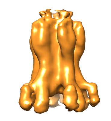 emd_4562.png emd_4562.png | 91.1 KB | ||
| Archive directory |  http://ftp.pdbj.org/pub/emdb/structures/EMD-4562ftp://ftp.pdbj.org/pub/emdb/structures/EMD-4562 http://ftp.pdbj.org/pub/emdb/structures/EMD-4562ftp://ftp.pdbj.org/pub/emdb/structures/EMD-4562 | HTTPS FTP |
-Related structure data
| Related structure data |  0264C  0265C  0266C  0267C  4561C  6hs7C C: citing same article ( |
|---|---|
| Similar structure data |

-Links
| EMDB pages | EMDB (EBI/PDBe) / EMDataResource |
|---|
-Map
| File | Download / File: emd_4562.map.gz / Format: CCP4 / Size: 530.3 KB / Type: IMAGE STORED AS FLOATING POINT NUMBER (4 BYTES) | ||||||||||||||||||||||||||||||||||||||||||||||||||||||||||||||||||||
|---|---|---|---|---|---|---|---|---|---|---|---|---|---|---|---|---|---|---|---|---|---|---|---|---|---|---|---|---|---|---|---|---|---|---|---|---|---|---|---|---|---|---|---|---|---|---|---|---|---|---|---|---|---|---|---|---|---|---|---|---|---|---|---|---|---|---|---|---|---|
| Projections & slices | Image control
Images are generated by Spider. generated in cubic-lattice coordinate | ||||||||||||||||||||||||||||||||||||||||||||||||||||||||||||||||||||
| Voxel size | X=Y=Z: 6.898 Å | ||||||||||||||||||||||||||||||||||||||||||||||||||||||||||||||||||||
| Density |
| ||||||||||||||||||||||||||||||||||||||||||||||||||||||||||||||||||||
| Symmetry | Space group: 1 | ||||||||||||||||||||||||||||||||||||||||||||||||||||||||||||||||||||
| Details | EMDB XML:
CCP4 map header:
| ||||||||||||||||||||||||||||||||||||||||||||||||||||||||||||||||||||
 Z (Sec.)
 Y (Row.)
 X (Col.)


-Supplemental data
- Sample components
-Entire : Membrane complex of the type 6 secretion system
| Entire | Name: Membrane complex of the type 6 secretion system |
|---|---|
| Components |
|
-Supramolecule #1: Membrane complex of the type 6 secretion system
| Supramolecule | Name: Membrane complex of the type 6 secretion system / type: complex / ID: 1 / Parent: 0 |
|---|---|
| Source (natural) | Organism: |
| Recombinant expression | Organism: |
| Molecular weight | Experimental: 1.7 MDa |
-Experimental details
-Structure determination
| Method | cryo EM |
|---|---|
 Processing Processing | subtomogram averaging |
| Aggregation state | cell |
-Sample preparation
| Buffer | pH: 7 |
|---|---|
| Vitrification | Cryogen name: ETHANE-PROPANE |
- Electron microscopy
| Microscope | FEI TITAN KRIOS |
|---|---|
| Specialist optics | Phase plate: VOLTA PHASE PLATE / Energy filter - Name: GIF Quantum LS / Energy filter - Slit width: 30 eV |
| Image recording | Film or detector model: GATAN K2 SUMMIT (4k x 4k) / Detector mode: COUNTING / Average exposure time: 3.0 sec. / Average electron dose: 1.3 e/Å2 Details: STA from various tomograms taken with slightly different electron dose and frame |
| Electron beam | Acceleration voltage: 300 kV / Electron source:  FIELD EMISSION GUN FIELD EMISSION GUN |
| Electron optics | Illumination mode: FLOOD BEAM / Imaging mode: BRIGHT FIELD |
| Experimental equipment |  Model: Titan Krios / Image courtesy: FEI Company |
-Image processing
| Final reconstruction | Applied symmetry - Point group: C5 (5 fold cyclic) / Algorithm: BACK PROJECTION / Resolution.type: BY AUTHOR / Resolution: 25.0 Å / Resolution method: FSC 0.5 CUT-OFF / Software - Name: PEET / Number subtomograms used: 23500 |
|---|---|
| Extraction | Number tomograms: 16 / Number images used: 28463 / Reference model: one single particle |
| Final angle assignment | Type: OTHER |
