 ムービー
ムービー コントローラー


+ データを開く

- 基本情報
基本情報
| 登録情報 | データベース: EMDB / ID: EMD-13878 | ||||||||||||
|---|---|---|---|---|---|---|---|---|---|---|---|---|---|
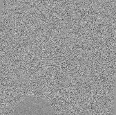
| タイトル | Cryo-electron tomogram from a cryo-FIB lift-out lamella of Drosophila melanogaster egg chambers | ||||||||||||
 マップデータ マップデータ | Tomogram from cryo-lift-out of D. melanogaster egg chambers | ||||||||||||
 試料 試料 |
| ||||||||||||
 キーワード キーワード | cyro-FIB / cryo-electron tomography / D. melanogaster / Golgi apparatus / RIBOSOME | ||||||||||||
| 生物種 |  | ||||||||||||
| 手法 | 電子線トモグラフィー法 / クライオ電子顕微鏡法 | ||||||||||||
 データ登録者 データ登録者 | Klumpe S / Fung HKH / Goetz SK / Plitzko JM / Mahamid J | ||||||||||||
| 資金援助 | European Union, 3件
| ||||||||||||
 引用 引用 | ジャーナル: Elife / 年: 2021 タイトル: A modular platform for automated cryo-FIB workflows. 著者: Sven Klumpe / Herman Kh Fung / Sara K Goetz / Ievgeniia Zagoriy / Bernhard Hampoelz / Xiaojie Zhang / Philipp S Erdmann / Janina Baumbach / Christoph W Müller / Martin Beck / Jürgen M Plitzko / Julia Mahamid /  要旨: Lamella micromachining by focused ion beam milling at cryogenic temperature (cryo-FIB) has matured into a preparation method widely used for cellular cryo-electron tomography. Due to the limited ...Lamella micromachining by focused ion beam milling at cryogenic temperature (cryo-FIB) has matured into a preparation method widely used for cellular cryo-electron tomography. Due to the limited ablation rates of low Ga ion beam currents required to maintain the structural integrity of vitreous specimens, common preparation protocols are time-consuming and labor intensive. The improved stability of new-generation cryo-FIB instruments now enables automated operations. Here, we present an open-source software tool, SerialFIB, for creating automated and customizable cryo-FIB preparation protocols. The software encompasses a graphical user interface for easy execution of routine lamellae preparations, a scripting module compatible with available Python packages, and interfaces with three-dimensional correlative light and electron microscopy (CLEM) tools. SerialFIB enables the streamlining of advanced cryo-FIB protocols such as multi-modal imaging, CLEM-guided lamella preparation and in situ lamella lift-out procedures. Our software therefore provides a foundation for further development of advanced cryogenic imaging and sample preparation protocols. | ||||||||||||
| 履歴 |
|
- 構造の表示
構造の表示
| ムービー |
 ムービービューア ムービービューア |
|---|---|
| 添付画像 |
- ダウンロードとリンク
-EMDBアーカイブ
| マップデータ | emd_13878.map.gz | 711.4 MB | EMDBマップデータ形式 | |
|---|---|---|---|---|
| ヘッダ (付随情報) | emd-13878-v30.xmlemd-13878.xml | 9.9 KB 9.9 KB | 表示 表示 | EMDBヘッダ |
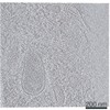
| 画像 |  emd_13878.png emd_13878.png | 259 KB | ||
| Filedesc metadata | emd-13878.cif.gz | 3.8 KB | ||
| アーカイブディレクトリ |  http://ftp.pdbj.org/pub/emdb/structures/EMD-13878ftp://ftp.pdbj.org/pub/emdb/structures/EMD-13878 http://ftp.pdbj.org/pub/emdb/structures/EMD-13878ftp://ftp.pdbj.org/pub/emdb/structures/EMD-13878 | HTTPS FTP |
-検証レポート
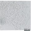
| 文書・要旨 | emd_13878_validation.pdf.gz | 569.1 KB | 表示 | EMDB検証レポート |
|---|---|---|---|---|
| 文書・詳細版 | emd_13878_full_validation.pdf.gz | 568.6 KB | 表示 | |
| XML形式データ | emd_13878_validation.xml.gz | 4.8 KB | 表示 | |
| CIF形式データ | emd_13878_validation.cif.gz | 5.3 KB | 表示 | |
| アーカイブディレクトリ | https://ftp.pdbj.org/pub/emdb/validation_reports/EMD-13878ftp://ftp.pdbj.org/pub/emdb/validation_reports/EMD-13878 | HTTPS FTP |
-関連構造データ


-リンク
| EMDBのページ | EMDB (EBI/PDBe) / EMDataResource |
|---|
-マップ
| ファイル | ダウンロード / ファイル: emd_13878.map.gz / 形式: CCP4 / 大きさ: 768.7 MB / タイプ: IMAGE STORED AS FLOATING POINT NUMBER (4 BYTES) | ||||||||||||||||||||||||||||||||||||||||||||||||||||||||||||||||||||
|---|---|---|---|---|---|---|---|---|---|---|---|---|---|---|---|---|---|---|---|---|---|---|---|---|---|---|---|---|---|---|---|---|---|---|---|---|---|---|---|---|---|---|---|---|---|---|---|---|---|---|---|---|---|---|---|---|---|---|---|---|---|---|---|---|---|---|---|---|---|
| 注釈 | Tomogram from cryo-lift-out of D. melanogaster egg chambers | ||||||||||||||||||||||||||||||||||||||||||||||||||||||||||||||||||||
| ボクセルのサイズ | X=Y=Z: 14.08 Å | ||||||||||||||||||||||||||||||||||||||||||||||||||||||||||||||||||||
| 密度 |
| ||||||||||||||||||||||||||||||||||||||||||||||||||||||||||||||||||||
| 対称性 | 空間群: 1 | ||||||||||||||||||||||||||||||||||||||||||||||||||||||||||||||||||||
| 詳細 | EMDB XML:
CCP4マップ ヘッダ情報:
| ||||||||||||||||||||||||||||||||||||||||||||||||||||||||||||||||||||
-添付データ
- 試料の構成要素
-全体 : D. melanogaster
| 全体 | 名称: D. melanogaster |
|---|---|
| 要素 |
|
-超分子 #1: D. melanogaster
| 超分子 | 名称: D. melanogaster / タイプ: tissue / ID: 1 / 親要素: 0 詳細: Tomogram from cryo-FIB lift-out lamella performed on a D. melanogaster egg chamber |
|---|---|
| 由来(天然) | 生物種: 株: w[*]; P{w[+mC]=sqh-mCherry.M}3 (Flybase ID: FBst0059024) 組織: Egg Chamber |
-実験情報
-構造解析
| 手法 | クライオ電子顕微鏡法 |
|---|---|
 解析 解析 | 電子線トモグラフィー法 |
| 試料の集合状態 | tissue |
-試料調製
| 緩衝液 | pH: 7.4 |
|---|---|
| 凍結 | 凍結剤: NITROGEN / 詳細: Leica EM Ice. |
| 加圧凍結法 | 装置: OTHER 詳細: 20% ficol in Schneider's medium. The value given for _em_high_pressure_freezing.instrument is EM Ice. This is not in a list of allowed values {'LEICA EM HPM100', 'LEICA EM PACT', 'BAL-TEC HPM ...詳細: 20% ficol in Schneider's medium. The value given for _em_high_pressure_freezing.instrument is EM Ice. This is not in a list of allowed values {'LEICA EM HPM100', 'LEICA EM PACT', 'BAL-TEC HPM 010', 'LEICA EM PACT2', 'EMS-002 RAPID IMMERSION FREEZER', 'OTHER'} so OTHER is written into the XML file. |
| Cryo protectant | Ficol |
| 切片作成 | 集束イオンビーム - 装置: OTHER / 集束イオンビーム - イオン: OTHER / 集束イオンビーム - 電圧: 30 / 集束イオンビーム - 電流: 1 / 集束イオンビーム - 時間: 60 / 集束イオンビーム - 温度: 93 K / 集束イオンビーム - Initial thickness: 10000 / 集束イオンビーム - 最終 厚さ: 200 集束イオンビーム - 詳細: Cryo-FIB Lift-Out method Various milling steps. The value given for _em_focused_ion_beam.instrument is TFS Aquilos. This is not in a list of allowed values ...集束イオンビーム - 詳細: Cryo-FIB Lift-Out method Various milling steps. The value given for _em_focused_ion_beam.instrument is TFS Aquilos. This is not in a list of allowed values {'DB235', 'OTHER'} so OTHER is written into the XML file. |
- 電子顕微鏡法
| 顕微鏡 | FEI TITAN KRIOS |
|---|---|
| 特殊光学系 | エネルギーフィルター - 名称: GIF Quantum LS / エネルギーフィルター - スリット幅: 20 eV |
| 撮影 | フィルム・検出器のモデル: GATAN K2 QUANTUM (4k x 4k) 検出モード: COUNTING / 平均電子線量: 2.0 e/Å2 |
| 電子線 | 加速電圧: 300 kV / 電子線源:  FIELD EMISSION GUN FIELD EMISSION GUN |
| 電子光学系 | 照射モード: FLOOD BEAM / 撮影モード: BRIGHT FIELD / Cs: 2.7 mm |
| 実験機器 |  モデル: Titan Krios / 画像提供: FEI Company |
-画像解析
| 最終 再構成 | ソフトウェア - 名称: IMOD / 使用した粒子像数: 59 |
|---|
