ムービー
ムービー コントローラー


+ データを開く

- 基本情報
基本情報
| 登録情報 | データベース: EMDB / ID: EMD-3068 | |||||||||
|---|---|---|---|---|---|---|---|---|---|---|
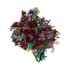
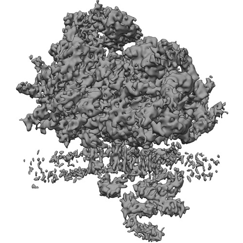
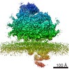
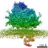
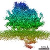
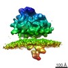
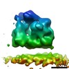
| タイトル | Mammalian ribosome bound to the native Sec61 protein-conducting channel in the 'non-inserting' state ('conventional' alignment) | |||||||||
 マップデータ マップデータ | Subtomogram average of non-solubilized ribosome-Sec61 complexes in the 'non-inserting' state. Subtomogram alignment was carried out using 'conventional' alignment. | |||||||||
 試料 試料 |
| |||||||||
 キーワード キーワード |  Ribosome (リボソーム) / Sec61 (Sec61) / Translocon (トランスロコン) / Endoplasmic Reticulum (小胞体) / Cryoelectron Tomography / Subtomogram Analysis Ribosome (リボソーム) / Sec61 (Sec61) / Translocon (トランスロコン) / Endoplasmic Reticulum (小胞体) / Cryoelectron Tomography / Subtomogram Analysis | |||||||||
| 機能・相同性 |  機能・相同性情報 機能・相同性情報Insertion of tail-anchored proteins into the endoplasmic reticulum membrane / membrane docking / endoplasmic reticulum Sec complex / pronephric nephron development / cotranslational protein targeting to membrane / Ssh1 translocon complex / Sec61 translocon complex / protein targeting to ER / protein-transporting ATPase activity / protein insertion into ER membrane ...Insertion of tail-anchored proteins into the endoplasmic reticulum membrane / membrane docking / endoplasmic reticulum Sec complex / pronephric nephron development / cotranslational protein targeting to membrane / Ssh1 translocon complex / Sec61 translocon complex / protein targeting to ER / protein-transporting ATPase activity / protein insertion into ER membrane / SRP-dependent cotranslational protein targeting to membrane, translocation / signal sequence binding / post-translational protein targeting to membrane, translocation / protein transmembrane transporter activity / phospholipid binding / ribosome binding / endoplasmic reticulum membrane / 小胞体 / 生体膜類似検索 - 分子機能 | |||||||||
| 生物種 |  Canis lupus familiaris (イヌ) Canis lupus familiaris (イヌ) | |||||||||
| 手法 | サブトモグラム平均法 / クライオ電子顕微鏡法 / 解像度: 9.0 Å | |||||||||
 データ登録者 データ登録者 | Pfeffer S / Burbaum L / Unverdorben P / Pech M / Chen Y / Zimmermann R / Beckmann R / Foerster F | |||||||||
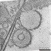
 引用 引用 | ジャーナル: Nat Commun / 年: 2015 タイトル: Structure of the native Sec61 protein-conducting channel. 著者: Stefan Pfeffer / Laura Burbaum / Pia Unverdorben / Markus Pech / Yuxiang Chen / Richard Zimmermann / Roland Beckmann / Friedrich Förster /  要旨: In mammalian cells, secretory and membrane proteins are translocated across or inserted into the endoplasmic reticulum (ER) membrane by the universally conserved protein-conducting channel Sec61, ...In mammalian cells, secretory and membrane proteins are translocated across or inserted into the endoplasmic reticulum (ER) membrane by the universally conserved protein-conducting channel Sec61, which has been structurally studied in isolated, detergent-solubilized states. Here we structurally and functionally characterize native, non-solubilized ribosome-Sec61 complexes on rough ER vesicles using cryo-electron tomography and ribosome profiling. Surprisingly, the 9-Å resolution subtomogram average reveals Sec61 in a laterally open conformation, even though the channel is not in the process of inserting membrane proteins into the lipid bilayer. In contrast to recent mechanistic models for polypeptide translocation and insertion, our results indicate that the laterally open conformation of Sec61 is the only conformation present in the ribosome-bound translocon complex, independent of its functional state. Consistent with earlier functional studies, our structure suggests that the ribosome alone, even without a nascent chain, is sufficient for lateral opening of Sec61 in a lipid environment. | |||||||||
| 履歴 |
|
- 構造の表示
構造の表示
| ムービー |
ムービービューア |
|---|---|
| 構造ビューア | EMマップ: SurfViewMolmilJmol/JSmol |
| 添付画像 |
- ダウンロードとリンク
-EMDBアーカイブ
| マップデータ | emd_3068.map.gz | 6.9 MB | EMDBマップデータ形式 | |
|---|---|---|---|---|
| ヘッダ (付随情報) | emd-3068-v30.xmlemd-3068.xml | 11.1 KB 11.1 KB | 表示 表示 | EMDBヘッダ |
| 画像 | EMD-3068.tif | 209.8 KB | ||
| アーカイブディレクトリ |  http://ftp.pdbj.org/pub/emdb/structures/EMD-3068ftp://ftp.pdbj.org/pub/emdb/structures/EMD-3068 http://ftp.pdbj.org/pub/emdb/structures/EMD-3068ftp://ftp.pdbj.org/pub/emdb/structures/EMD-3068 | HTTPS FTP |
-関連構造データ
-リンク
| EMDBのページ | EMDB (EBI/PDBe) / EMDataResource |
|---|---|
| 「今月の分子」の関連する項目 |


-マップ
| ファイル | ダウンロード / ファイル: emd_3068.map.gz / 形式: CCP4 / 大きさ: 39.7 MB / タイプ: IMAGE STORED AS FLOATING POINT NUMBER (4 BYTES) | ||||||||||||||||||||||||||||||||||||||||||||||||||||||||||||
|---|---|---|---|---|---|---|---|---|---|---|---|---|---|---|---|---|---|---|---|---|---|---|---|---|---|---|---|---|---|---|---|---|---|---|---|---|---|---|---|---|---|---|---|---|---|---|---|---|---|---|---|---|---|---|---|---|---|---|---|---|---|
| 注釈 | Subtomogram average of non-solubilized ribosome-Sec61 complexes in the 'non-inserting' state. Subtomogram alignment was carried out using 'conventional' alignment. | ||||||||||||||||||||||||||||||||||||||||||||||||||||||||||||
| ボクセルのサイズ | X=Y=Z: 2.62 Å | ||||||||||||||||||||||||||||||||||||||||||||||||||||||||||||
| 密度 |
| ||||||||||||||||||||||||||||||||||||||||||||||||||||||||||||
| 対称性 | 空間群: 1 | ||||||||||||||||||||||||||||||||||||||||||||||||||||||||||||
| 詳細 | EMDB XML:
CCP4マップ ヘッダ情報:
| ||||||||||||||||||||||||||||||||||||||||||||||||||||||||||||
-添付データ
- 試料の構成要素
-全体 : Mammalian ribosome bound to the native protein translocon on cani...
| 全体 | 名称: Mammalian ribosome bound to the native protein translocon on canine pancreatic ER vesicles |
|---|---|
| 要素 |
|
-超分子 #1000: Mammalian ribosome bound to the native protein translocon on cani...
| 超分子 | 名称: Mammalian ribosome bound to the native protein translocon on canine pancreatic ER vesicles タイプ: sample / ID: 1000 / Number unique components: 2 |
|---|
-超分子 #1: Membrane-bound 80S ribosome
| 超分子 | 名称: Membrane-bound 80S ribosome / タイプ: complex / ID: 1 / 組換発現: No / Ribosome-details: ribosome-eukaryote: ALL |
|---|---|
| 由来(天然) | 生物種: Canis lupus familiaris (イヌ) / 別称: Dog / 組織: Pancreas |
-分子 #1: ER protein translocon
| 分子 | 名称: ER protein translocon / タイプ: protein_or_peptide / ID: 1 / 組換発現: No |
|---|---|
| 由来(天然) | 生物種: Canis lupus familiaris (イヌ) / 別称: Dog / 組織: Pancreas / Organelle: Endoplasmic Reticulum |
-実験情報
-構造解析
| 手法 | クライオ電子顕微鏡法 |
|---|---|
 解析 解析 | サブトモグラム平均法 |
| 試料の集合状態 | particle |
-試料調製
| 濃度 | 2 mg/mL |
|---|---|
| 緩衝液 | pH: 7.6 / 詳細: 20mM Hepes, 50mM KCl; 2mM MgCl2 |
| グリッド | 詳細: Lacey carbon molybdenum grid |
| 凍結 | 凍結剤: ETHANE-PROPANE MIXTURE / チャンバー内湿度: 70 % / 装置: FEI VITROBOT MARK IV / 手法: Blot 3 seconds before plunging. |
- 電子顕微鏡法
| 顕微鏡 | FEI TITAN KRIOS |
|---|---|
| 電子線 | 加速電圧: 300 kV / 電子線源: FIELD EMISSION GUN |
| 電子光学系 | 照射モード: FLOOD BEAM / 撮影モード: BRIGHT FIELDBright-field microscopy / 最大 デフォーカス(公称値): 4.0 µm / 最小 デフォーカス(公称値): 3.0 µm |
| 特殊光学系 | エネルギーフィルター - 名称: Gatan |
| 試料ステージ | 試料ホルダーモデル: FEI TITAN KRIOS AUTOGRID HOLDER Tilt series - Axis1 - Min angle: -20 ° / Tilt series - Axis1 - Max angle: 20 ° |
| 日付 | 2014年6月18日 |
| 撮影 | カテゴリ: CCD / フィルム・検出器のモデル: GATAN K2 (4k x 4k) / 平均電子線量: 30 e/Å2 |
| 実験機器 |  モデル: Titan Krios / 画像提供: FEI Company |
-画像解析
| CTF補正 | 詳細: Each tilt image |
|---|---|
| 最終 再構成 | 想定した対称性 - 点群: C1 (非対称) / 解像度のタイプ: BY AUTHOR / 解像度: 9.0 Å / 解像度の算出法: OTHER ソフトウェア - 名称: PyTom, tom_toolbox, av3_toolbox 使用したサブトモグラム数: 17600 |
| 詳細 | Tomogram reconstruction and template matching against a single particle cryo-EM reconstruction of the 80S ribosome were accomplished using PyTom. Subtomograms extracted from cross correlation peaks in the tomogram were classified using constrained principal component analysis focusing on the large ribosomal subunit and the ER membrane. For the retained coordinates, 1 x binned subtomograms were reconstructed individually from the weighted back-projections using the full tilt range, iteratively aligned and classified focusing on the translocon. For the retained coordinates, unbinned subtomograms were reconstructed individually from the weighted back-projections using only a reduced tilt range (-20 deg to +20 deg) and iteratively aligned using a 'conventional' subtomogram alignment procedure. |
-原子モデル構築 1
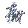
| 初期モデル | PDB ID: Chain - #0 - Chain ID: 1 / Chain - #1 - Chain ID: 2 / Chain - #2 - Chain ID: 3 |
|---|---|
| ソフトウェア | 名称: MDFF |
| 詳細 | The Sec61a N-terminal domain along with Sec61b was first fitted as a rigid body prior to flexible fitting. |
| 精密化 | 空間: REAL / プロトコル: FLEXIBLE FIT / 当てはまり具合の基準: pseudo-energy |
| 得られたモデル | 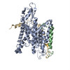 PDB-5a6u: |