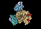
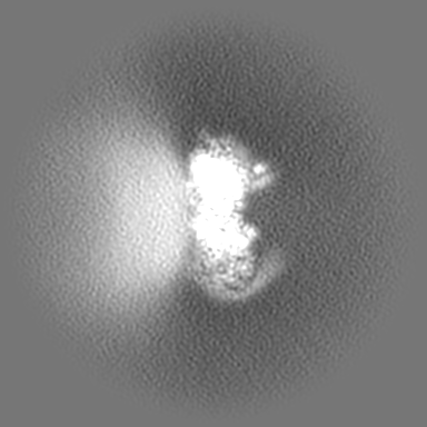
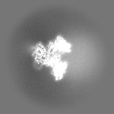
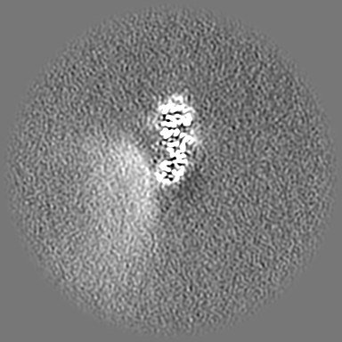
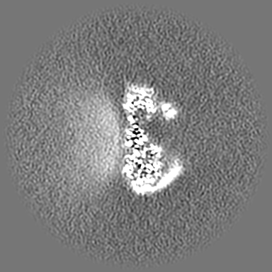
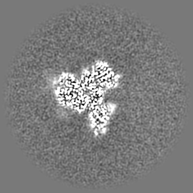
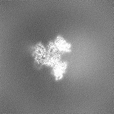
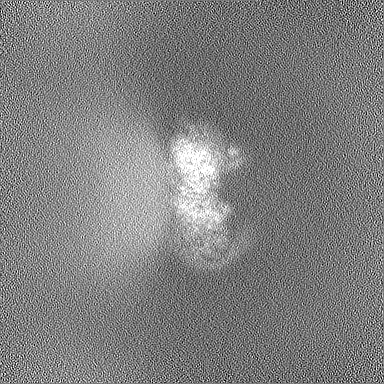
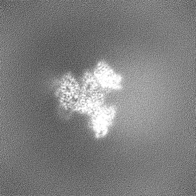
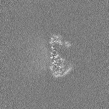
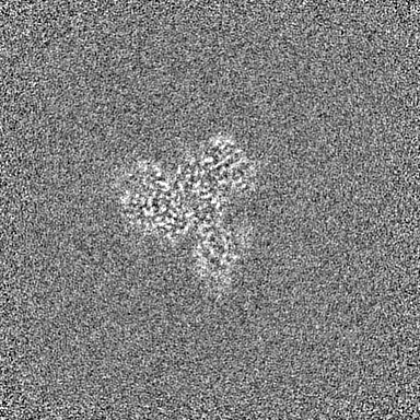
登録情報 データベース : EMDB / ID : EMD-28268タイトル Phospholipase C beta 3 (PLCb3) in complex with Gbg on lipid nanodiscs final sharpened map 複合体 : Phospholipase C beta 3 (PLCb3) in complex with two copies of Gbg on lipid nanodiscs.複合体 : Phospholipase C beta 3 (PLCb3)タンパク質・ペプチド : 1-phosphatidylinositol 4,5-bisphosphate phosphodiesterase beta-3複合体 : G protein beta subunit (Gb1)タンパク質・ペプチド : Guanine nucleotide-binding protein G(I)/G(S)/G(T) subunit beta-1複合体 : G protein gamma subunit 2 (Gg2)タンパク質・ペプチド : Guanine nucleotide-binding protein G(I)/G(S)/G(O) subunit gamma-2複合体 : G protein beta subunit (Gb1)複合体 : G protein gamma subunit 2 (Gg2)リガンド : CALCIUM ION機能・相同性 分子機能 ドメイン・相同性 構成要素
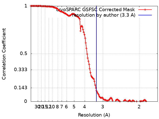
/ / / / / / / / / / / / / / / / / / / / / / / / / / / / / / / / / / / / / / / / / / / / / / / / / / / / / / / / / / / / / / / / / / / / / / / / / / / / / / / / / / / / / / / / / / / / / / / / / / / / / / / / / / / / / / / / / / / / / / / / / / / / / / / / / / / / / / / / / / / / / 生物種 Homo sapiens (ヒト)手法 / / 解像度 : 3.3 Å Falzone ME / MacKinnon R 資金援助 Organization Grant number 国 Howard Hughes Medical Institute (HHMI) National Institutes of Health/National Institute of General Medical Sciences (NIH/NIGMS) F32GM142137
ジャーナル : Proc Natl Acad Sci U S A / 年 : 2023タイトル : activates hydrolysis by recruiting and orienting on the membrane surface.著者 : Maria E Falzone / Roderick MacKinnon / 要旨 : catalyze the hydrolysis of phosphatidylinositol 4, 5-bisphosphate [Formula: see text] into [Formula: see text] [Formula: see text] and [Formula: see text] [Formula: see text]. [Formula: see text] ... catalyze the hydrolysis of phosphatidylinositol 4, 5-bisphosphate [Formula: see text] into [Formula: see text] [Formula: see text] and [Formula: see text] [Formula: see text]. [Formula: see text] regulates the activity of many membrane proteins, while and lead to increased intracellular Ca levels and activate protein kinase C, respectively. are regulated by G protein-coupled receptors through direct interaction with [Formula: see text] and [Formula: see text] and are aqueous-soluble enzymes that must bind to the cell membrane to act on their lipid substrate. This study addresses the mechanism by which [Formula: see text] activates 3. We show that 3 functions as a slow Michaelis-Menten enzyme ( [Formula: see text] ) on membrane surfaces. We used membrane partitioning experiments to study the solution-membrane localization equilibrium of 3. Its partition coefficient is such that only a small quantity of 3 exists in the membrane in the absence of [Formula: see text] . When [Formula: see text] is present, equilibrium binding on the membrane surface increases 3 in the membrane, increasing [Formula: see text] in proportion. Atomic structures on membrane vesicle surfaces show that two [Formula: see text] anchor 3 with its catalytic site oriented toward the membrane surface. Taken together, the enzyme kinetic, membrane partitioning, and structural data show that [Formula: see text] activates by increasing its concentration on the membrane surface and orienting its catalytic core to engage [Formula: see text] . This principle of activation explains rapid stimulated catalysis with low background activity, which is essential to the biological processes mediated by [Formula: see text], and . 履歴 登録 2022年9月28日 - ヘッダ(付随情報) 公開 2023年5月24日 - マップ公開 2023年5月24日 - 更新 2023年5月24日 - 現状 2023年5月24日 処理サイト : RCSB / 状態 : 公開
すべて表示 表示を減らす
 ムービー
ムービー コントローラー

 万見
万見 データを開く
 基本情報
基本情報
 マップデータ
 試料
試料 機能・相同性情報
 phosphoinositide phospholipase C / Fatty Acids bound to GPR40 (FFAR1) regulate insulin secretion / Acetylcholine regulates insulin secretion / phosphatidylinositol metabolic process / PLC beta mediated events / regulation of systemic arterial blood pressure / phosphatidylinositol phospholipase C activity /
 データ登録者
 米国, 2件
  引用
引用 構造の表示
構造の表示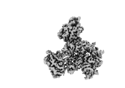
 ダウンロードとリンク
 emd_28268.png
 http://ftp.pdbj.org/pub/emdb/structures/EMD-28268


 Z
Z Y
Y X
X


 試料の構成要素
 解析
解析 電子顕微鏡法