Movie
Movie Controller

[English] 日本語
 Yorodumi
- PDB-8ear: Structure of the full-length IP3R1 channel determined in the pres... -
+ Open data

- Basic information
| Entry | Database: PDB / ID: 8ear | ||||||||||||||||||||||||
|---|---|---|---|---|---|---|---|---|---|---|---|---|---|---|---|---|---|---|---|---|---|---|---|---|---|
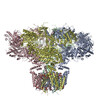
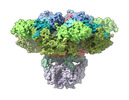
| Title | Structure of the full-length IP3R1 channel determined in the presence of Calcium/IP3/ATP | ||||||||||||||||||||||||
 Components Components | Inositol 1,4,5-trisphosphate receptor type 1 | ||||||||||||||||||||||||
 Keywords Keywords |  MEMBRANE PROTEIN / Calcium channel / lipid nanodisc MEMBRANE PROTEIN / Calcium channel / lipid nanodisc | ||||||||||||||||||||||||
| Function / homology |  Function and homology information Function and homology informationEffects of PIP2 hydrolysis / Antigen activates B Cell Receptor (BCR) leading to generation of second messengers / inositol 1,4,5-trisphosphate receptor activity involved in regulation of postsynaptic cytosolic calcium levels / Elevation of cytosolic Ca2+ levels / cGMP effects / smooth endoplasmic reticulum membrane / platelet dense tubular network / negative regulation of calcium-mediated signaling / calcineurin complex / platelet dense granule membrane ...Effects of PIP2 hydrolysis / Antigen activates B Cell Receptor (BCR) leading to generation of second messengers / inositol 1,4,5-trisphosphate receptor activity involved in regulation of postsynaptic cytosolic calcium levels / Elevation of cytosolic Ca2+ levels / cGMP effects / smooth endoplasmic reticulum membrane / platelet dense tubular network / negative regulation of calcium-mediated signaling / calcineurin complex / platelet dense granule membrane / epithelial fluid transport / ion channel modulating, G protein-coupled receptor signaling pathway / phospholipase C-activating G protein-coupled acetylcholine receptor signaling pathway / inositol 1,4,5-trisphosphate-gated calcium channel activity / regulation of postsynaptic cytosolic calcium ion concentration / voluntary musculoskeletal movement / inositol 1,4,5 trisphosphate binding / positive regulation of calcium ion transport / Glucagon-like Peptide-1 (GLP1) regulates insulin secretion / endoplasmic reticulum calcium ion homeostasis / positive regulation of hepatocyte proliferation / nuclear inner membrane / transport vesicle membrane / Ion homeostasis / dendrite development / intracellularly gated calcium channel activity / ligand-gated ion channel signaling pathway / GABA-ergic synapse / intrinsic apoptotic signaling pathway in response to endoplasmic reticulum stress / calcium channel inhibitor activity / cellular response to cAMP / release of sequestered calcium ion into cytosol / phosphatidylinositol binding / post-embryonic development / secretory granule membrane / sarcoplasmic reticulum / synaptic membrane / liver regeneration / calcium-mediated signaling / calcium ion transmembrane transport / Schaffer collateral - CA1 synapse / cell morphogenesis / positive regulation of neuron projection development / positive regulation of insulin secretion / calcium ion transport / presynapse / nuclear envelope / phospholipase C-activating G protein-coupled receptor signaling pathway / positive regulation of cytosolic calcium ion concentration / cellular response to hypoxia / postsynapse / protein phosphatase binding / transmembrane transporter binding / postsynaptic density / response to hypoxia / positive regulation of apoptotic process / protein domain specific binding / dendrite / neuronal cell body / synapse / calcium ion binding / protein-containing complex binding / endoplasmic reticulum membrane / nucleolus / negative regulation of apoptotic process / perinuclear region of cytoplasm / endoplasmic reticulum / protein-containing complex / membrane / identical protein binding / plasma membrane / cytoplasmSimilarity search - Function | ||||||||||||||||||||||||
| Biological species |  Rattus norvegicus (Norway rat) Rattus norvegicus (Norway rat) | ||||||||||||||||||||||||
| Method | ELECTRON MICROSCOPY / single particle reconstruction / cryo EM / Resolution: 3.5 Å | ||||||||||||||||||||||||
 Authors Authors | Fan, G. / Baker, M.R. / Terry, L.E. / Arige, V. / Chen, M. / Seryshev, A.B. / Baker, M.L. / Ludtke, S.J. / Yule, D.I. / Serysheva, I.I. | ||||||||||||||||||||||||
| Funding support |  United States, 7items United States, 7items
| ||||||||||||||||||||||||
 Citation Citation | Journal: Nat Commun / Year: 2022 Title: Conformational motions and ligand-binding underlying gating and regulation in IPR channel. Authors: Guizhen Fan / Mariah R Baker / Lara E Terry / Vikas Arige / Muyuan Chen / Alexander B Seryshev / Matthew L Baker / Steven J Ludtke / David I Yule / Irina I Serysheva / Abstract: Inositol-1,4,5-trisphosphate receptors (IPRs) are activated by IP and Ca and their gating is regulated by various intracellular messengers that finely tune the channel activity. Here, using single ...Inositol-1,4,5-trisphosphate receptors (IPRs) are activated by IP and Ca and their gating is regulated by various intracellular messengers that finely tune the channel activity. Here, using single particle cryo-EM analysis we determined 3D structures of the nanodisc-reconstituted IPR1 channel in two ligand-bound states. These structures provide unprecedented details governing binding of IP, Ca and ATP, revealing conformational changes that couple ligand-binding to channel opening. Using a deep-learning approach and 3D variability analysis we extracted molecular motions of the key protein domains from cryo-EM density data. We find that IP binding relies upon intrinsic flexibility of the ARM2 domain in the tetrameric channel. Our results highlight a key role of dynamic side chains in regulating gating behavior of IPR channels. This work represents a stepping-stone to developing mechanistic understanding of conformational pathways underlying ligand-binding, activation and regulation of the channel. | ||||||||||||||||||||||||
| History |
|
- Structure visualization
| Structure viewer | Molecule: MolmilJmol/JSmol |
|---|
- Downloads & links
-Download
| PDBx/mmCIF format | 8ear.cif.gz | 1.7 MB | Display | PDBx/mmCIF format |
|---|---|---|---|---|
| PDB format | pdb8ear.ent.gz | 1.4 MB | Display | PDB format |
| PDBx/mmJSON format | 8ear.json.gz | Tree view | PDBx/mmJSON format | |
| Others |  Other downloads Other downloads |
-Validation report
| Arichive directory | https://data.pdbj.org/pub/pdb/validation_reports/ea/8earftp://data.pdbj.org/pub/pdb/validation_reports/ea/8ear | HTTPS FTP |
|---|
-Related structure data
| Related structure data | 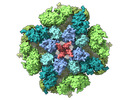 27983MC 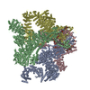 8eaqC M: map data used to model this data C: citing same article ( |
|---|---|
| Similar structure data |
-Links
 PDBj
PDBj


- Assembly
| Deposited unit | 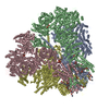
|
|---|---|
| 1 |
|
-Components
-Protein , 1 types, 4 molecules ABCD
| #1: Protein | Mass: 313657.406 Da / Num. of mol.: 4 / Source method: isolated from a natural source / Source: (natural) Rattus norvegicus (Norway rat) / Organ: cerebellum / References: UniProt: P29994 |
|---|
-Non-polymers , 5 types, 56 molecules 

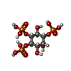

| #2: Chemical | ChemComp-ZN /  Mass: 65.409 Da / Num. of mol.: 4 / Source method: obtained synthetically / Formula: Zn Mass: 65.409 Da / Num. of mol.: 4 / Source method: obtained synthetically / Formula: Zn#3: Chemical | ChemComp-ATP / Adenosine triphosphate Mass: 507.181 Da / Num. of mol.: 4 / Source method: obtained synthetically / Formula: C10H16N5O13P3 / Feature type: SUBJECT OF INVESTIGATION / Comment: ATP, energy-carrying molecule*YM Mass: 507.181 Da / Num. of mol.: 4 / Source method: obtained synthetically / Formula: C10H16N5O13P3 / Feature type: SUBJECT OF INVESTIGATION / Comment: ATP, energy-carrying molecule*YM#4: Chemical | ChemComp-I3P / Inositol trisphosphate Mass: 420.096 Da / Num. of mol.: 4 / Source method: obtained synthetically / Formula: C6H15O15P3 / Feature type: SUBJECT OF INVESTIGATION Mass: 420.096 Da / Num. of mol.: 4 / Source method: obtained synthetically / Formula: C6H15O15P3 / Feature type: SUBJECT OF INVESTIGATION#5: Chemical | ChemComp-CA /  Mass: 40.078 Da / Num. of mol.: 16 / Source method: obtained synthetically / Formula: Ca / Feature type: SUBJECT OF INVESTIGATION Mass: 40.078 Da / Num. of mol.: 16 / Source method: obtained synthetically / Formula: Ca / Feature type: SUBJECT OF INVESTIGATION#6: Chemical | ChemComp-PLX / (  Mass: 767.132 Da / Num. of mol.: 28 / Source method: obtained synthetically / Formula: C42H89NO8P / Comment: phospholipid*YM Mass: 767.132 Da / Num. of mol.: 28 / Source method: obtained synthetically / Formula: C42H89NO8P / Comment: phospholipid*YM |
|---|
-Details
| Has ligand of interest | Y |
|---|
-Experimental details
-Experiment
| Experiment | Method: ELECTRON MICROSCOPY |
|---|---|
| EM experiment | Aggregation state: PARTICLE / 3D reconstruction method: single particle reconstruction |
- Sample preparation
| Component | Name: Type 1 inositol 1,4,5-trisphosphate receptor tetrameric protein complex Type: COMPLEX / Entity ID: #1 / Source: NATURAL |
|---|---|
| Molecular weight | Value: 1.3 MDa / Experimental value: NO |
| Source (natural) | Organism: Rattus norvegicus (Norway rat) / Cellular location: membrane / Organ: cerebellum / Organelle: endoplasmic reticulum |
| Buffer solution | pH: 7.4 |
| Specimen | Conc.: 1 mg/ml / Embedding applied: NO / Shadowing applied: NO / Staining applied: NO / Vitrification applied: YES / Details: reconstituted in lipid nanodisc |
| Specimen support | Grid material: COPPER / Grid mesh size: 200 divisions/in. / Grid type: Quantifoil R2/1 |
| Vitrification | Instrument: FEI VITROBOT MARK IV / Cryogen name: ETHANE / Humidity: 100 % / Chamber temperature: 277 K |
- Electron microscopy imaging
| Experimental equipment |  Model: Titan Krios / Image courtesy: FEI Company |
|---|---|
| Microscopy | Model: TFS KRIOS |
| Electron gun | Electron source: FIELD EMISSION GUN / Accelerating voltage: 300 kV / Illumination mode: FLOOD BEAM |
| Electron lens | Mode: BRIGHT FIELDBright-field microscopy / Nominal magnification: 130000 X / Calibrated magnification: 46943 X / Nominal defocus max: 2500 nm / Nominal defocus min: 800 nm / Cs: 2.7 mm / Alignment procedure: COMA FREE |
| Specimen holder | Cryogen: NITROGEN / Specimen holder model: FEI TITAN KRIOS AUTOGRID HOLDER |
| Image recording | Average exposure time: 0.2 sec. / Electron dose: 49 e/Å2 / Detector mode: SUPER-RESOLUTION / Film or detector model: GATAN K2 QUANTUM (4k x 4k) / Num. of real images: 24478 |
| EM imaging optics | Energyfilter name: GIF Bioquantum / Energyfilter slit width: 20 eV |
| Image scans | Sampling size: 5 µm / Width: 3840 / Height: 3712 / Movie frames/image: 35 |
- Processing
| Software |
| ||||||||||||||||||||||||||||||||||||
|---|---|---|---|---|---|---|---|---|---|---|---|---|---|---|---|---|---|---|---|---|---|---|---|---|---|---|---|---|---|---|---|---|---|---|---|---|---|
| EM software |
| ||||||||||||||||||||||||||||||||||||
| CTF correction | Type: NONE | ||||||||||||||||||||||||||||||||||||
| Particle selection | Num. of particles selected: 1452797 / Details: NeuralNet autopicking in EMAN2 | ||||||||||||||||||||||||||||||||||||
| Symmetry | Point symmetry: C4 (4 fold cyclic) | ||||||||||||||||||||||||||||||||||||
| 3D reconstruction | Resolution: 3.5 Å / Resolution method: FSC 0.143 CUT-OFF / Num. of particles: 133740 / Symmetry type: POINT | ||||||||||||||||||||||||||||||||||||
| Atomic model building | Protocol: FLEXIBLE FIT / Space: REAL | ||||||||||||||||||||||||||||||||||||
| Atomic model building | PDB-ID: 7LHE | ||||||||||||||||||||||||||||||||||||
| Refine LS restraints |
|