 Movie
Movie Controller


+ Open data

- Basic information
| Entry | Database: PDB / ID: 1ayl | ||||||
|---|---|---|---|---|---|---|---|
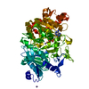
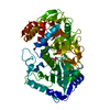
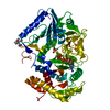
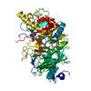
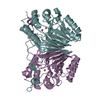
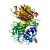
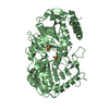
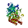
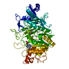
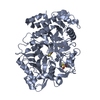
| Title | PHOSPHOENOLPYRUVATE CARBOXYKINASE | ||||||
 Components Components | PHOSPHOENOLPYRUVATE CARBOXYKINASE | ||||||
 Keywords Keywords | KINASE (TRANSPHOSPHORYLATING) / P-LOOP / PROTEIN-ATP COMPLEX / NUCLEOTIDE-TRIPHOSPHATE HYDROLASE | ||||||
| Function / homology |  Function and homology informationphosphoenolpyruvate carboxykinase (ATP) / phosphoenolpyruvate carboxykinase (ATP) activity / gluconeogenesis / calcium ion binding / magnesium ion binding / ATP binding / cytosol Function and homology informationphosphoenolpyruvate carboxykinase (ATP) / phosphoenolpyruvate carboxykinase (ATP) activity / gluconeogenesis / calcium ion binding / magnesium ion binding / ATP binding / cytosolSimilarity search - Function | ||||||
| Biological species |  Escherichia coli (E. coli) Escherichia coli (E. coli) | ||||||
| Method | X-RAY DIFFRACTION / SYNCHROTRON / Resolution: 1.8 Å | ||||||
 Authors Authors | Tari, L.W. / Pugazenthi, U. / Goldie, H. / Delbaere, L.T.J. | ||||||
 Citation Citation | Journal: Nat.Struct.Biol. / Year: 1996 Title: Snapshot of an enzyme reaction intermediate in the structure of the ATP-Mg2+-oxalate ternary complex of Escherichia coli PEP carboxykinase. Authors: Tari, L.W. / Matte, A. / Pugazhenthi, U. / Goldie, H. / Delbaere, L.T. #1: Journal: To be PublishedTitle: Allosteric Control by Calcium and Mechanism of Desensitization of Phosphoenolpyruvate Carboxykinase of Escherichia Coli Authors: Goldie, H. / Sanwal, B.D. #2: Journal: J.Mol.Biol. / Year: 1996Title: Crystal Structure of Escherichia Coli Phosphoenolpyruvate Carboxykinase: A New Structural Family with the P-Loop Nucleoside Triphosphate Hydrolase Fold Authors: Matte, A. / Goldie, H. / Sweet, R.M. / Delbaere, L.T. #3: Journal: J.Mol.Biol. / Year: 1991Title: Crystallization of the Calcium-Activated Phosphoenolpyruvate Carboxykinase from Escherichia Coli K12 Authors: Delbaere, L.T. / Vandonselaar, M. / Glaeske, D. / Jabs, C. / Goldie, H. #4: Journal: J.Bacteriol. / Year: 1990Title: Sequence of the Pcka Gene of Escherichia Coli K-12: Relevance to Genetic and Allosteric Regulation and Homology of E. Coli Phosphoenolpyruvate Carboxykinase with the Enzymes from Trypanosoma ...Title: Sequence of the Pcka Gene of Escherichia Coli K-12: Relevance to Genetic and Allosteric Regulation and Homology of E. Coli Phosphoenolpyruvate Carboxykinase with the Enzymes from Trypanosoma Brucei and Saccharomyces Cerevisiae Authors: Medina, V. / Pontarollo, R. / Glaeske, D. / Tabel, H. / Goldie, H. | ||||||
| History |
|
- Structure visualization
| Structure viewer | Molecule: MolmilJmol/JSmol |
|---|
- Downloads & links
-Download
| PDBx/mmCIF format | 1ayl.cif.gz | 155.1 KB | Display | PDBx/mmCIF format |
|---|---|---|---|---|
| PDB format | pdb1ayl.ent.gz | 121 KB | Display | PDB format |
| PDBx/mmJSON format | 1ayl.json.gz | Tree view | PDBx/mmJSON format | |
| Others |  Other downloads Other downloads |
-Validation report
| Arichive directory | https://data.pdbj.org/pub/pdb/validation_reports/ay/1aylftp://data.pdbj.org/pub/pdb/validation_reports/ay/1ayl | HTTPS FTP |
|---|
-Related structure data
| Similar structure data |
|---|
-Links
 PDBj
PDBj- Assembly
| Deposited unit | 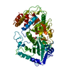
| ||||||||
|---|---|---|---|---|---|---|---|---|---|
| 1 |
| ||||||||
| Unit cell |
|
-Components
| #1: Protein | / ATP-OXALOACETATE CARBOXY-LYASE (ATP) Mass: 59796.254 Da / Num. of mol.: 1 / Source method: isolated from a natural source / Details: ORDERED MAGNESIUM ION OBSERVED BOUND TO ATP / Source: (natural) Escherichia coli (E. coli) / Strain: K12References: UniProt: P22259, phosphoenolpyruvate carboxykinase (ATP) |
|---|---|
| #2: Chemical | ChemComp-OXL / Oxalate  Mass: 88.019 Da / Num. of mol.: 1 / Source method: obtained synthetically / Formula: C2O4 Mass: 88.019 Da / Num. of mol.: 1 / Source method: obtained synthetically / Formula: C2O4 |
| #3: Chemical | ChemComp-MG /   Mass: 24.305 Da / Num. of mol.: 1 / Source method: obtained synthetically / Formula: Mg Mass: 24.305 Da / Num. of mol.: 1 / Source method: obtained synthetically / Formula: Mg |
| #4: Chemical | ChemComp-ATP / Adenosine triphosphate  Mass: 507.181 Da / Num. of mol.: 1 / Source method: obtained synthetically / Formula: C10H16N5O13P3 / Comment: ATP, energy-carrying molecule*YM Mass: 507.181 Da / Num. of mol.: 1 / Source method: obtained synthetically / Formula: C10H16N5O13P3 / Comment: ATP, energy-carrying molecule*YM |
| #5: Water | ChemComp-HOH / Water Mass: 18.015 Da / Num. of mol.: 345 / Source method: isolated from a natural source / Formula: H2O Mass: 18.015 Da / Num. of mol.: 345 / Source method: isolated from a natural source / Formula: H2O |
-Experimental details
-Experiment
| Experiment | Method: X-RAY DIFFRACTION |
|---|
- Sample preparation
| Crystal | Density Matthews: 2.4 Å3/Da / Density % sol: 49 % | ||||||||||||||||||||||||||||||||||||||||||||||||||||||||||||||||||||||||
|---|---|---|---|---|---|---|---|---|---|---|---|---|---|---|---|---|---|---|---|---|---|---|---|---|---|---|---|---|---|---|---|---|---|---|---|---|---|---|---|---|---|---|---|---|---|---|---|---|---|---|---|---|---|---|---|---|---|---|---|---|---|---|---|---|---|---|---|---|---|---|---|---|---|
| Crystal | *PLUS | ||||||||||||||||||||||||||||||||||||||||||||||||||||||||||||||||||||||||
| Crystal grow | *PLUS Temperature: 21 ℃ / pH: 4.4 / Method: vapor diffusion, hanging drop | ||||||||||||||||||||||||||||||||||||||||||||||||||||||||||||||||||||||||
| Components of the solutions | *PLUS
|
-Data collection
| Diffraction source | Source: SYNCHROTRON / Site: Photon Factory  / Beamline: BL-6B / Wavelength: 1 / Beamline: BL-6B / Wavelength: 1 |
|---|---|
| Detector | Type: FUJI / Detector: IMAGE PLATE |
| Radiation | Monochromatic (M) / Laue (L): M / Scattering type: x-ray |
| Radiation wavelength | Wavelength: 1 Å / Relative weight: 1 |
| Reflection | Num. obs: 40067 / % possible obs: 76.7 % / Observed criterion σ(I): 1 / Redundancy: 2.2 % / Rmerge(I) obs: 0.076 |
| Reflection | *PLUS Highest resolution: 1.8 Å |
- Processing
| Software |
| ||||||||||||||||||||||||||||||||||||||||||||||||||||||||||||
|---|---|---|---|---|---|---|---|---|---|---|---|---|---|---|---|---|---|---|---|---|---|---|---|---|---|---|---|---|---|---|---|---|---|---|---|---|---|---|---|---|---|---|---|---|---|---|---|---|---|---|---|---|---|---|---|---|---|---|---|---|---|
| Refinement | Resolution: 1.8→6 Å / σ(F): 2 Details: MET 477 HAS DISALLOWED RAMACHANDRAN ANGLES; THE DEPOSITORS ACCOUNT FOR THIS AS IT IS THE SECOND RESIDUE WITHIN A GAMMA-TURN.
| ||||||||||||||||||||||||||||||||||||||||||||||||||||||||||||
| Displacement parameters | Biso mean: 21.42 Å2 | ||||||||||||||||||||||||||||||||||||||||||||||||||||||||||||
| Refine analyze | Luzzati coordinate error obs: 0.2 Å | ||||||||||||||||||||||||||||||||||||||||||||||||||||||||||||
| Refinement step | Cycle: LAST / Resolution: 1.8→6 Å
| ||||||||||||||||||||||||||||||||||||||||||||||||||||||||||||
| Refine LS restraints |
| ||||||||||||||||||||||||||||||||||||||||||||||||||||||||||||
| Software | *PLUS Name: X-PLOR / Classification: refinement | ||||||||||||||||||||||||||||||||||||||||||||||||||||||||||||
| Refine LS restraints | *PLUS
|
