+ データを開く
- 基本情報
基本情報
| 登録情報 | データベース: SASBDB / ID: SASDBY2 |
|---|---|
 試料 試料 | Inorganic pyrophosphatase (PPase) from E. coli
|
| 機能・相同性 |  機能・相同性情報 機能・相同性情報inorganic triphosphate phosphatase activity / inorganic diphosphatase / inorganic diphosphate phosphatase activity / phosphate-containing compound metabolic process / magnesium ion binding / zinc ion binding / membrane / cytosol 類似検索 - 分子機能 |
| 生物種 |  |
 引用 引用 |  ジャーナル: PLoS One / 年: 2016 ジャーナル: PLoS One / 年: 2016タイトル: X-Ray Solution Scattering Study of Four Escherichia coli Enzymes Involved in Stationary-Phase Metabolism. 著者: Liubov A Dadinova / Eleonora V Shtykova / Petr V Konarev / Elena V Rodina / Natalia E Snalina / Natalia N Vorobyeva / Svetlana A Kurilova / Tatyana I Nazarova / Cy M Jeffries / Dmitri I Svergun /   要旨: The structural analyses of four metabolic enzymes that maintain and regulate the stationary growth phase of Escherichia coli have been performed primarily drawing on the results obtained from ...The structural analyses of four metabolic enzymes that maintain and regulate the stationary growth phase of Escherichia coli have been performed primarily drawing on the results obtained from solution small angle X-ray scattering (SAXS) and other structural techniques. The proteins are (i) class I fructose-1,6-bisphosphate aldolase (FbaB); (ii) inorganic pyrophosphatase (PPase); (iii) 5-keto-4-deoxyuronate isomerase (KduI); and (iv) glutamate decarboxylase (GadA). The enzyme FbaB, that until now had an unknown structure, is predicted to fold into a TIM-barrel motif that form globular protomers which SAXS experiments show associate into decameric assemblies. In agreement with previously reported crystal structures, PPase forms hexamers in solution that are similar to the previously reported X-ray crystal structure. Both KduI and GadA that are responsible for carbohydrate (pectin) metabolism and acid stress responses, respectively, form polydisperse mixtures consisting of different oligomeric states. Overall the SAXS experiments yield additional insights into shape and organization of these metabolic enzymes and further demonstrate the utility of hybrid methods, i.e., solution SAXS combined with X-ray crystallography, bioinformatics and predictive 3D-structural modeling, as tools to enrich structural studies. The results highlight the structural complexity that the protein components of metabolic networks may adopt which cannot be fully captured using individual structural biology techniques. |
 登録者 登録者 |
|
- 構造の表示
構造の表示
| 構造ビューア | 分子:  Molmil Molmil Jmol/JSmol Jmol/JSmol |
|---|
- ダウンロードとリンク
-モデル
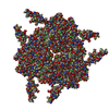
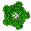
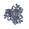
| モデル #418 | 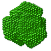 タイプ: dummy / ダミー原子の半径: 2.00 A / 対称性: P32 / カイ2乗値: 1.032  Omokage検索でこの集合体の類似形状データを探す (詳細) Omokage検索でこの集合体の類似形状データを探す (詳細) |
|---|---|
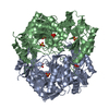
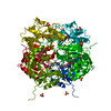
| モデル #419 |  タイプ: atomic / ダミー原子の半径: 1.90 A / 対称性: P32 / カイ2乗値: 1.437  Omokage検索でこの集合体の類似形状データを探す (詳細) Omokage検索でこの集合体の類似形状データを探す (詳細) |
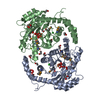
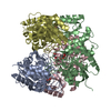
| モデル #420 | 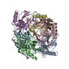 タイプ: atomic / カイ2乗値: 5.180  Omokage検索でこの集合体の類似形状データを探す (詳細) Omokage検索でこの集合体の類似形状データを探す (詳細) |
- 試料
試料
 試料 試料 | 名称: Inorganic pyrophosphatase (PPase) from E. coli / 試料濃度: 1.40-10.80 |
|---|---|
| バッファ | 名称: 50 mM Tris 10 mM NaCl / 濃度: 50.00 mM / pH: 7.5 / 組成: 10 mM NaCl |
| 要素 #279 | 名称: PPase / タイプ: protein / 記述: Inorganic pyrophosphatase (PPase) from E. coli / 分子量: 19.56 / 分子数: 6 / 由来: Escherichia coli / 参照: UniProt: P0A7A9 配列: SLLNVPAGKD LPEDIYVVIE IPANADPIKY EIDKESGALF VDRFMSTAMF YPCNYGYINH TLSLDGDPVD VLVPTPYPLQ PGSVTRCRPV GVLKMTDEAG EDAKLVAVPH SKLSKEYDHI KDVNDLPELL KAQIAHFFEH YKDLEKGKWV KVEGWENAEA AKAEIVASFE RAKNK |
-実験情報
| ビーム | 設備名称: PETRA III EMBL P12 / 地域: Hamburg / 国: Germany  / 線源: X-ray synchrotron / 波長: 0.12 Å / スペクトロメータ・検出器間距離: 3.1 mm / 線源: X-ray synchrotron / 波長: 0.12 Å / スペクトロメータ・検出器間距離: 3.1 mm | ||||||||||||||||||||||||||||||||||||
|---|---|---|---|---|---|---|---|---|---|---|---|---|---|---|---|---|---|---|---|---|---|---|---|---|---|---|---|---|---|---|---|---|---|---|---|---|---|
| 検出器 | 名称: Pilatus 2M | ||||||||||||||||||||||||||||||||||||
| スキャン | 測定日: 2013年6月30日 / 保管温度: 10 °C / セル温度: 10 °C / 照射時間: 0.05 sec. / フレーム数: 20 / 単位: 1/nm /
| ||||||||||||||||||||||||||||||||||||
| 距離分布関数 P(R) |
| ||||||||||||||||||||||||||||||||||||
| 結果 | コメント: The models displayed above are, from top to bottom: 1) An individual DAMMIN dummy atom model; 2) SASREF rigid body model with the associated CRYSOL30 fit to the data and; 3) The PPase ...コメント: The models displayed above are, from top to bottom: 1) An individual DAMMIN dummy atom model; 2) SASREF rigid body model with the associated CRYSOL30 fit to the data and; 3) The PPase crystal structure fit to the data (derived from Protein data bank entry 2AUU). All of the individual DAMMIN models, associated fits and averaging can be found in the zip archive for this entry.
|
 ムービー
ムービー コントローラー


 SASDBY2