+ データを開く
- 基本情報
基本情報
| 登録情報 | データベース: SASBDB / ID: SASDFN8 |
|---|---|
 試料 試料 | Apoferritin from horse spleen - SEC-SAXS coupled to multiangle laser and quasi-elastic light scattering (MALLS and QELS)
|
| 機能・相同性 |  機能・相同性情報 機能・相同性情報ferritin complex / autolysosome / ferric iron binding / autophagosome / iron ion transport / ferrous iron binding / cytoplasmic vesicle / intracellular iron ion homeostasis / iron ion binding / cytoplasm 類似検索 - 分子機能 |
| 生物種 |  |
 登録者 登録者 |
|
- 構造の表示
構造の表示
| 構造ビューア | 分子:  Molmil Molmil Jmol/JSmol Jmol/JSmol |
|---|
- ダウンロードとリンク
-モデル
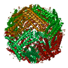
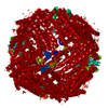
| モデル #3208 | 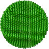 タイプ: dummy / ソフトウェア: (SUPCOMB 23 (r9988)) / ダミー原子の半径: 2.70 A / 対称性: P1 / カイ2乗値: 1.074 / P-value: 0.577890  Omokage検索でこの集合体の類似形状データを探す (詳細) Omokage検索でこの集合体の類似形状データを探す (詳細) |
|---|---|
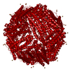
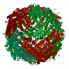
| モデル #3209 |  タイプ: dummy / ソフトウェア: (DAMFILT 5.0 (r10552)) / ダミー原子の半径: 4.00 A / カイ2乗値: 1.074 / P-value: 0.577890  Omokage検索でこの集合体の類似形状データを探す (詳細) Omokage検索でこの集合体の類似形状データを探す (詳細) |
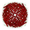
| モデル #3210 | 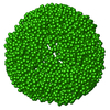 タイプ: dummy / ダミー原子の半径: 1.90 A / 対称性: P432 / カイ2乗値: 5.88  Omokage検索でこの集合体の類似形状データを探す (詳細) Omokage検索でこの集合体の類似形状データを探す (詳細) |
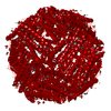
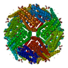
| モデル #3212 | 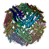 タイプ: atomic / ソフトウェア: (PDB) / カイ2乗値: 8.846  Omokage検索でこの集合体の類似形状データを探す (詳細) Omokage検索でこの集合体の類似形状データを探す (詳細) |
- 試料
試料
 試料 試料 | 名称: Apoferritin from horse spleen - SEC-SAXS coupled to multiangle laser and quasi-elastic light scattering (MALLS and QELS) 試料濃度: 11 mg/ml |
|---|---|
| バッファ | 名称: 50 mM HEPES, 150 mM NaCl, 2% v/v glycerol, / pH: 7 / コメント: Running buffer for SEC-SAXS |
| 要素 #1766 | タイプ: protein / 記述: Apoferritin light chain / 分子量: 19.977 / 分子数: 24 / 由来: Equus caballus / 参照: UniProt: P02791 配列: MSSQIRQNYS TEVEAAVNRL VNLYLRASYT YLSLGFYFDR DDVALEGVCH FFRELAEEKR EGAERLLKMQ NQRGGRALFQ DLQKPSQDEW GTTLDAMKAA IVLEKSLNQA LLDLHALGSA QADPHLCDFL ESHFLDEEVK LIKKMGDHLT NIQRLVGSQA GLGEYLFERL TLKHD |
-実験情報
| ビーム | 設備名称: PETRA III EMBL P12 / 地域: Hamburg / 国: Germany  / 線源: X-ray synchrotron / 波長: 0.123982 Å / スペクトロメータ・検出器間距離: 3 mm / 線源: X-ray synchrotron / 波長: 0.123982 Å / スペクトロメータ・検出器間距離: 3 mm | ||||||||||||||||||||||||||||||
|---|---|---|---|---|---|---|---|---|---|---|---|---|---|---|---|---|---|---|---|---|---|---|---|---|---|---|---|---|---|---|---|
| 検出器 | 名称: Pilatus 6M | ||||||||||||||||||||||||||||||
| スキャン | 測定日: 2019年4月5日 / 保管温度: 20 °C / セル温度: 20 °C / 照射時間: 1 sec. / フレーム数: 85 / 単位: 1/nm /
| ||||||||||||||||||||||||||||||
| 距離分布関数 P(R) |
| ||||||||||||||||||||||||||||||
| 結果 | コメント: Apoferritin underwent pre-purification prior SEC-SAXS using the following method. All procedures were performed at 4 oC. An apoferritin stock solution (from equine spleen supplied in ca. ...コメント: Apoferritin underwent pre-purification prior SEC-SAXS using the following method. All procedures were performed at 4 oC. An apoferritin stock solution (from equine spleen supplied in ca. 50% v/v glycerol; Sigma Gel Filtration Markers Kit MWGF1000) was diluted two-fold in 25 mM HEPES, 50 mM NaCl, 5 mM urea, 1% v/v glycerol, pH 7. Approximately 200 μl of sample were loaded onto a Superose 6 Increase 10/300 column (GE Healthcare) equilibrated in the same buffer (flow rate = 0.4 ml/min). Fractionated aliquots corresponding to the highest absorbing peak (estimated using UV A280 and UV A245 nm) were pooled and concentrated (30 kDa centrifuge spin filter) to a final concentration of 11 mg/ml (the concentration was determined from triplicate UV A280 measurements using an E0.1% of 0.729 (= 1 g/l) calculated from the amino acid sequence (ProtParam)). Approximately 50 μl aliquots were snap-frozen in liquid nitrogen then stored at -80 oC prior to the SEC-SAXS analysis that was performed at room temperature in 50 mM HEPES, 150 mM NaCl, 2% v/v glycerol, pH 7. The Rg-correlation through the SEC-SAXS peak, the individual unsubtracted SEC-SAXS frames as well as the results from coupled MALLS and QELS analysis are included in the full entry zip archive. The quoted experimental molecular weight was determined using MALLS in combination with refractive-index (RI) measurements that were recorded from the same sample eluting from the column using a split-flow SEC-SAXS-light scattering configuration (Graewert et al., (2015) Sci. Reports. 5, 10734: doi: 10.1038/srep10734). The average hydrodynamic radius of the protein is 6.7 nm.
|
 ムービー
ムービー コントローラー


 SASDFN8