 Movie
Movie Controller


+ Open data

- Basic information
| Entry | Database: PDB / ID: 2ffx | ||||||
|---|---|---|---|---|---|---|---|
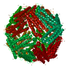
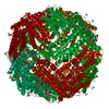
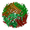
| Title | Structure of Human Ferritin L. Chain | ||||||
 Components Components | ferritin light chain | ||||||
 Keywords Keywords | METAL BINDING PROTEIN / Human Ferritin Light Chain Perdeuterated Capsid | ||||||
| Function / homology |  Function and homology information Function and homology informationferritin complex / Scavenging by Class A Receptors / Golgi Associated Vesicle Biogenesis / autolysosome / ferric iron binding / autophagosome / iron ion transport / ferrous iron binding / Iron uptake and transport / azurophil granule lumen ...ferritin complex / Scavenging by Class A Receptors / Golgi Associated Vesicle Biogenesis / autolysosome / ferric iron binding / autophagosome / iron ion transport / ferrous iron binding / Iron uptake and transport / azurophil granule lumen / intracellular iron ion homeostasis / iron ion binding / Neutrophil degranulation / extracellular exosome / extracellular region / membrane / identical protein binding / cytoplasm / cytosol Similarity search - Function | ||||||
| Biological species |  Homo sapiens (human) Homo sapiens (human) | ||||||
| Method |  X-RAY DIFFRACTION / SYNCHROTRON / MOLECULAR REPLACEMENT / Resolution: 1.9 Å X-RAY DIFFRACTION / SYNCHROTRON / MOLECULAR REPLACEMENT / Resolution: 1.9 Å | ||||||
 Authors Authors | Wang, Z.M. / Li, C. / Ellenburg, M.P. / Soitsman, E.M. / Ruble, J.R. / Wright, B.S. / Ho, J.X. / Carter, D.C. | ||||||
 Citation Citation | Journal: ACTA CRYSTALLOGR.,SECT.D / Year: 2006 Title: Structure of human ferritin L chain Authors: Wang, Z.M. / Li, C. / Ellenburg, M.P. / Soistman, E.M. / Ruble, J.R. / Wright, B.S. / Ho, J.X. / Carter, D.C. | ||||||
| History |
|
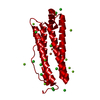
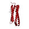
- Structure visualization
| Structure viewer | Molecule: MolmilJmol/JSmol |
|---|
- Downloads & links
-Download
| PDBx/mmCIF format | 2ffx.cif.gz | 53.9 KB | Display | PDBx/mmCIF format |
|---|---|---|---|---|
| PDB format | pdb2ffx.ent.gz | 39.2 KB | Display | PDB format |
| PDBx/mmJSON format | 2ffx.json.gz | Tree view | PDBx/mmJSON format | |
| Others |  Other downloads Other downloads |
-Validation report
| Arichive directory | https://data.pdbj.org/pub/pdb/validation_reports/ff/2ffxftp://data.pdbj.org/pub/pdb/validation_reports/ff/2ffx | HTTPS FTP |
|---|
-Related structure data


-Links
 PDBj
PDBj


- Assembly
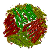
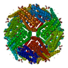
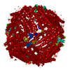
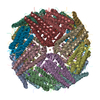
| Deposited unit | 
| ||||||||||||
|---|---|---|---|---|---|---|---|---|---|---|---|---|---|
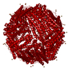
| 1 | x 24
| ||||||||||||
| Unit cell |
| ||||||||||||
| Components on special symmetry positions |
|
-Components
| #1: Protein | Mass: 19802.396 Da / Num. of mol.: 1 Source method: isolated from a genetically manipulated source Source: (gene. exp.) Homo sapiens (human) / Description: perdeuterated salts and solvent / Gene: FTL / Plasmid: pET 11a-LF / Production host:  | ||||
|---|---|---|---|---|---|
| #2: Chemical | ChemComp-CD /   Mass: 112.411 Da / Num. of mol.: 14 / Source method: obtained synthetically / Formula: Cd Mass: 112.411 Da / Num. of mol.: 14 / Source method: obtained synthetically / Formula: Cd#3: Chemical | ChemComp-SO4 / |   Mass: 96.063 Da / Num. of mol.: 1 / Source method: obtained synthetically / Formula: SO4 Mass: 96.063 Da / Num. of mol.: 1 / Source method: obtained synthetically / Formula: SO4#4: Water | ChemComp-HOH / |  Mass: 18.015 Da / Num. of mol.: 155 / Source method: isolated from a natural source / Formula: H2O Mass: 18.015 Da / Num. of mol.: 155 / Source method: isolated from a natural source / Formula: H2O |
-Experimental details
-Experiment
| Experiment | Method: X-RAY DIFFRACTION / Number of used crystals: 1 |
|---|
- Sample preparation
| Crystal | Density Matthews: 3.66 Å3/Da / Density % sol: 66.44 % |
|---|---|
| Crystal grow | Temperature: 290 K / Method: vapor diffusion / pH: 5.5 Details: 1.6% Cadmium Sulfate, 0.2M Sodium Acetate, pH 5.5, VAPOR DIFFUSION, temperature 290K |
-Data collection
| Diffraction | Mean temperature: 100 K |
|---|---|
| Diffraction source | Source: SYNCHROTRON / Site: NSLS  / Beamline: X12B / Wavelength: 1 Å / Beamline: X12B / Wavelength: 1 Å |
| Detector | Type: ADSC QUANTUM 315 / Detector: CCD / Date: Nov 22, 2003 |
| Radiation | Protocol: SINGLE WAVELENGTH / Monochromatic (M) / Laue (L): M / Scattering type: x-ray |
| Radiation wavelength | Wavelength: 1 Å / Relative weight: 1 |
| Reflection | Resolution: 1.9→50 Å / Num. all: 23698 / Num. obs: 23698 / % possible obs: 100 % / Redundancy: 5.3 % / Biso Wilson estimate: 19.7 Å2 / Rsym value: 0.1 / Net I/σ(I): 7.4 |
- Processing
| Software |
| ||||||||||||||||||||||||||||||||||||||||||||||||||||||||||||||||||||||||||||||||
|---|---|---|---|---|---|---|---|---|---|---|---|---|---|---|---|---|---|---|---|---|---|---|---|---|---|---|---|---|---|---|---|---|---|---|---|---|---|---|---|---|---|---|---|---|---|---|---|---|---|---|---|---|---|---|---|---|---|---|---|---|---|---|---|---|---|---|---|---|---|---|---|---|---|---|---|---|---|---|---|---|---|
| Refinement | Method to determine structure: MOLECULAR REPLACEMENT / Resolution: 1.9→43.76 Å / Rfactor Rfree error: 0.007 / Data cutoff high absF: 225145.68 / Data cutoff low absF: 0 / Isotropic thermal model: RESTRAINED / Cross valid method: THROUGHOUT / σ(F): 0 / Details: BULK SOLVENT MODEL USED
| ||||||||||||||||||||||||||||||||||||||||||||||||||||||||||||||||||||||||||||||||
| Solvent computation | Solvent model: FLAT MODEL / Bsol: 43.9903 Å2 / ksol: 0.372796 e/Å3 | ||||||||||||||||||||||||||||||||||||||||||||||||||||||||||||||||||||||||||||||||
| Displacement parameters | Biso mean: 23.8 Å2
| ||||||||||||||||||||||||||||||||||||||||||||||||||||||||||||||||||||||||||||||||
| Refine analyze |
| ||||||||||||||||||||||||||||||||||||||||||||||||||||||||||||||||||||||||||||||||
| Refinement step | Cycle: LAST / Resolution: 1.9→43.76 Å
| ||||||||||||||||||||||||||||||||||||||||||||||||||||||||||||||||||||||||||||||||
| Refine LS restraints |
| ||||||||||||||||||||||||||||||||||||||||||||||||||||||||||||||||||||||||||||||||
| LS refinement shell | Resolution: 1.9→2.02 Å / Rfactor Rfree error: 0.026 / Total num. of bins used: 6
| ||||||||||||||||||||||||||||||||||||||||||||||||||||||||||||||||||||||||||||||||
| Xplor file |
|
