 ムービー
ムービー コントローラー

+ データを開く

- 基本情報
基本情報
| 登録情報 | データベース: SASBDB / ID: SASDFS4 |
|---|---|
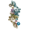
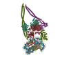
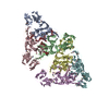
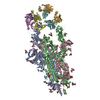
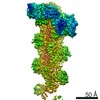
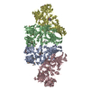
 試料 試料 | HP0242 from Helicobacter pylori, N-terminal domain of syntaxin-1A from Rattus norvegicus, de novo designed coiled-coil trimer domain (Domain A (PDB:3MLI)-Linker (PDB:1BR0)-Domain B (PDB:4DZL))
|
| 生物種 |   Helicobacter pylori (ピロリ菌) Helicobacter pylori (ピロリ菌) de novo design (unknown) |
 引用 引用 | ジャーナル: ACS Synth Biol / 年: 2019 タイトル: Construction of a Quadrangular Tetramer and a Cage-Like Hexamer from Three-Helix Bundle-Linked Fusion Proteins. 著者: Takaaki Miyamoto / Yugo Hayashi / Keito Yoshida / Hiroki Watanabe / Takayuki Uchihashi / Kento Yonezawa / Nobutaka Shimizu / Hironari Kamikubo / Shun Hirota /  要旨: Self-assembled protein nanostructures have gained interest, owing to their potential applications in biomaterials; however, successful design and construction of protein nanostructures are limited. ...Self-assembled protein nanostructures have gained interest, owing to their potential applications in biomaterials; however, successful design and construction of protein nanostructures are limited. Herein, we constructed fusion protein 1 by linking the C-terminus of a dimerization domain and the N-terminus of another dimerization domain with a three-helix bundle protein, where it self-assembled mainly into tetramers. By replacing the C-terminal dimerization domain of 1 with a trimerization domain (fusion protein 2), hexamers were mainly obtained. According to ab initio structural models reconstructed from the small-angle X-ray scattering data, the tetramer of 1 and hexamer of 2 adopted quadrangle and cage-like structures, respectively, although they were combinations of different conformations. High-speed atomic force microscopy observations indicated that the tetramer and hexamer exhibit conformational dynamics. These results show that the present method utilizing three-helix bundle-linked fusion proteins is useful in the construction of protein nanostructures. |
 登録者 登録者 |
|
- 構造の表示
構造の表示
| 構造ビューア | 分子:  MolmilJmol/JSmol MolmilJmol/JSmol |
|---|
- ダウンロードとリンク
 SASDFS4
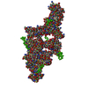


-モデル
| モデル #2930 |  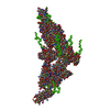 タイプ: mix / ダミー原子の半径: 1.90 A / カイ2乗値: 0.10 / P-value: 0.000088  Omokage検索でこの集合体の類似形状データを探す (詳細) Omokage検索でこの集合体の類似形状データを探す (詳細) |
|---|
-試料
| 試料 | 名称: HP0242 from Helicobacter pylori, N-terminal domain of syntaxin-1A from Rattus norvegicus, de novo designed coiled-coil trimer domain (Domain A (PDB:3MLI)-Linker (PDB:1BR0)-Domain B (PDB:4DZL)) 試料濃度: 1.90-6.80 |
|---|---|
| バッファ | 名称: 20 mM Tris-HCl 150 mM NaCl / pH: 8 |
| 要素 #1583 | 名称: Fusion protein2 / タイプ: protein 記述: HP0242 from Helicobacter pylori, N-terminal domain of syntaxin-1A from Rattus norvegicus, de novo designed coiled-coil trimer domain 分子量: 29.855 / 分子数: 6 由来: Helicobacter pylori, Rattus norvegicus, de novo design 配列: MGSSHHHHHH SSGLVPRGSH MRDYSELEIF EGNPLDKWND IIFHASKKAS KKELERLLEL LALCETFIEK EDLEEKFESF AKALRIDEEL QQKIESRKTD IVIQSMANIL SALFMDEFFE QVEEIRGFID KIAENVEEVK RKHSAILASP NPDEKTKEEL EELMSDIKKT ...配列: MGSSHHHHHH SSGLVPRGSH MRDYSELEIF EGNPLDKWND IIFHASKKAS KKELERLLEL LALCETFIEK EDLEEKFESF AKALRIDEEL QQKIESRKTD IVIQSMANIL SALFMDEFFE QVEEIRGFID KIAENVEEVK RKHSAILASP NPDEKTKEEL EELMSDIKKT ANKVRSKLKS IEQSIEQEEG LNRSSADLRI RKTQHSTLSR KFVEVMSEYN ATQSDYGSGG EIAAIKQEIA AIKKEIAAIK WEIAAIKQGY G |
-実験情報
| ビーム | 設備名称: Nara Institute of Science and Technology Rigaku Nano-Viewer 地域: Ikoma / 国: Japan / 線源: X-ray in house / 波長: 0.15418 Å / スペクトロメータ・検出器間距離: 0.75 mm | |||||||||||||||||||||||||||||||||||||||
|---|---|---|---|---|---|---|---|---|---|---|---|---|---|---|---|---|---|---|---|---|---|---|---|---|---|---|---|---|---|---|---|---|---|---|---|---|---|---|---|---|
| 検出器 | 名称: Pilatus 200K / タイプ: Pilatus / Pixsize x: 172 mm | |||||||||||||||||||||||||||||||||||||||
| スキャン |  測定日: 2017年3月29日 / 保管温度: 4 °C / セル温度: 20 °C / 照射時間: 60 sec. / フレーム数: 25 / 単位: 1/A /
| |||||||||||||||||||||||||||||||||||||||
| 距離分布関数 P(R) | 
| |||||||||||||||||||||||||||||||||||||||
| 結果 | 
|
