 Movie
Movie Controller
[English] 日本語
 Yorodumi
- SASDEC8: Bacillus thuringiensis LexA repressor bound to Bacteriophage pGIL... -
+ Open data

- Basic information
| Entry | Database: SASBDB / ID: SASDEC8 |
|---|---|
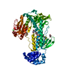
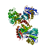
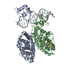
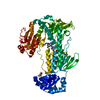
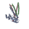
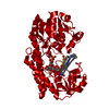
 Sample Sample | Bacillus thuringiensis LexA repressor bound to Bacteriophage pGIL01 gp7 (Bt_LexA_GIL01_gp7)
|
| Function / homology |  Function and homology information Function and homology informationrepressor LexA / SOS response / DNA replication / serine-type endopeptidase activity / DNA repair / negative regulation of DNA-templated transcription / proteolysis / DNA binding Similarity search - Function |
| Biological species |  Bacteriophage pGIL01 |
 Citation Citation | Date: 2019 May Title: Structural Insights into Bacteriophage GIL01 gp7 Inhibition of Host LexA Repressor Authors: Caveney N / Pavlin A / Caballero G / Bahun M / Hodnik V / de Castro L / Fornelos N / Butala M |
 Contact author Contact author |
|
- Structure visualization
| Structure viewer | Molecule: MolmilJmol/JSmol |
|---|
- Downloads & links
 SASDEC8
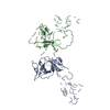


-Models
| Model #2676 |  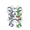 Type: atomic Comment: I-TASSER, ClusPro, ModLoop model of LexA-gp7 complex Chi-square value: 6.158  Search similar-shape structures of this assembly by Omokage search (details) Search similar-shape structures of this assembly by Omokage search (details) |
|---|---|
| Model #2677 |  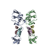 Type: atomic Comment: I-TASSER, ClusPro, ModLoop model of LexA-gp7 complex in SREFLEX, restrained Chi-square value: 2.580 / P-value: 0.000002 Search similar-shape structures of this assembly by Omokage search (details) |
| Model #2678 |   Type: atomic Comment: I-TASSER, ClusPro, ModLoop model of LexA-gp7 complex in SREFLEX, unrestrained Chi-square value: 1.899 / P-value: 0.000040 Search similar-shape structures of this assembly by Omokage search (details) |
-Sample
| Sample | Name: Bacillus thuringiensis LexA repressor bound to Bacteriophage pGIL01 gp7 (Bt_LexA_GIL01_gp7) Specimen concentration: 0.75-12.00 / Entity id: 1376 / 1377 |
|---|---|
| Buffer | Name: 20 mM Hepes, 300 mM NaCl, 10% glycerol, / pH: 8 |
| Entity #1376 | Name: LexA / Type: protein / Description: Bacillus thuringiensis LexA repressor / Formula weight: 23.394 / Num. of mol.: 2 / Source: Bacillus thuringiensis / References: UniProt: A0A2B4EEI3 Sequence: MLENMEKLTK RQQDILDFIK LKVQEKGYPP SVREIGQAVG LASSSTVHGH LSRLEEKGYI RRDPTKPRAI EILGEDRMDT ETQSVIQVPI VGKVTAGLPI TAVESVEEHF PLPASIVSGA DQVFMLRISG DSMIEAGIFD GDLVVVRQQQ SAYNGEIVVA LTEDNEATVK ...Sequence: MLENMEKLTK RQQDILDFIK LKVQEKGYPP SVREIGQAVG LASSSTVHGH LSRLEEKGYI RRDPTKPRAI EILGEDRMDT ETQSVIQVPI VGKVTAGLPI TAVESVEEHF PLPASIVSGA DQVFMLRISG DSMIEAGIFD GDLVVVRQQQ SAYNGEIVVA LTEDNEATVK RFYKEKDHFR LQPENSSLEP IILKQVSVIG KVIGVYRDLH |
| Entity #1377 | Name: gp7 / Type: protein / Description: Bacteriophage pGIL01 gp7 / Formula weight: 5.921 / Num. of mol.: 4 / Source: Bacteriophage pGIL01 / References: UniProt: Q7WSG2 Sequence: MRDKLLDFII ELSQSSKQVV SKSYVIDRLM QVTKEDYKEL EKNVEGKKDD |
-Experimental information
| Beam | Instrument name: University of British Columbia Rigaku BioSAXS-2000 City: Vancouver / 国: Canada  / Type of source: X-ray in house / Wavelength: 0.154 Å / Type of source: X-ray in house / Wavelength: 0.154 Å | ||||||
|---|---|---|---|---|---|---|---|
| Detector | Name: Rigaku HyOix-3000 / Type: Hybrid Pixel Array Detector / Pixsize x: 100 mm | ||||||
| Scan |  Measurement date: Aug 25, 2017 / Storage temperature: 6 °C / Cell temperature: 6 °C / Exposure time: 300 sec. / Number of frames: 12 / Unit: 1/A /
| ||||||
| Result | Experimental MW: 108.2 kDa / I0 from Guinier: 1.53 / Rg from Guinier: 4.391 nm / Type of curve: merged |
