 Movie
Movie Controller
[English] 日本語
 Yorodumi
- SASDDK9: Conformation of R1-3 human dystrophin fragment in interaction wit... -
+ Open data

- Basic information
| Entry | Database: SASBDB / ID: SASDDK9 |
|---|---|
 Sample Sample | Conformation of R1-3 human dystrophin fragment in interaction with zwitterionic phospholipid bicelles (SANS)
|
| Function / homology |  Function and homology information Function and homology informationregulation of muscle system process / regulation of cellular response to growth factor stimulus / syntrophin complex / cardiac muscle cell action potential / synaptic signaling / dystrophin-associated glycoprotein complex / cell-substrate junction / peptide biosynthetic process / motile cilium assembly / dystroglycan binding ...regulation of muscle system process / regulation of cellular response to growth factor stimulus / syntrophin complex / cardiac muscle cell action potential / synaptic signaling / dystrophin-associated glycoprotein complex / cell-substrate junction / peptide biosynthetic process / motile cilium assembly / dystroglycan binding / regulation of skeletal muscle contraction by regulation of release of sequestered calcium ion / vinculin binding / regulation of sodium ion transmembrane transport / costamere / Formation of the dystrophin-glycoprotein complex (DGC) / muscle cell development / regulation of calcium ion transmembrane transport / Striated Muscle Contraction / filopodium membrane / structural constituent of muscle / muscle organ development / muscle cell cellular homeostasis / myosin binding / maintenance of blood-brain barrier / neuron projection terminus / Non-integrin membrane-ECM interactions / nitric-oxide synthase binding / regulation of skeletal muscle contraction / neuron development / skeletal muscle tissue development / regulation of release of sequestered calcium ion into cytosol by sarcoplasmic reticulum / response to muscle stretch / cardiac muscle contraction / regulation of cardiac muscle contraction by regulation of the release of sequestered calcium ion / positive regulation of neuron differentiation / regulation of heart rate / filopodium / positive regulation of neuron projection development / sarcolemma / structural constituent of cytoskeleton / Z disc / intracellular protein localization / actin binding / protein-containing complex assembly / cytoskeleton / postsynaptic membrane / membrane raft / synapse / cell surface / protein-containing complex / zinc ion binding / nucleus / plasma membrane / cytosol Similarity search - Function |
| Biological species |  Homo sapiens (human) Homo sapiens (human) |
 Citation Citation | Date: 2018 Aug Title: Human dystrophin structural changes upon binding to anionic membrane lipids Authors: Santos Morais R / Delalande O / Pérez J / Mias-Lucquin D / Lagarrigue M / Martel A / Molza A / Chéron A / Raguénès-Nicol C / Chenuel T / Bondon A / Appavou M / Le Rumeur E / Combet S |
 Contact author Contact author |
|
- Structure visualization
| Structure viewer | Molecule: MolmilJmol/JSmol |
|---|
- Downloads & links
-Data source
| SASBDB page |  SASDDK9 SASDDK9 |
|---|
-Related structure data
| Related structure data | C: citing same article ( |
|---|---|
| Similar structure data |


-External links
| Related items in Molecule of the Month |
|---|


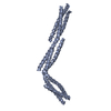
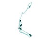
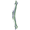
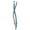
-Models
| Model #2140 |  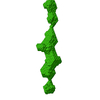 Type: dummy / Software: (2.7) / Radius of dummy atoms: 1.20 A / Chi-square value: 0.589 / P-value: 0.052545  Search similar-shape structures of this assembly by Omokage search (details) Search similar-shape structures of this assembly by Omokage search (details) |
|---|
-Sample
| Sample | Name: Conformation of R1-3 human dystrophin fragment in interaction with zwitterionic phospholipid bicelles (SANS) Specimen concentration: 4.2 mg/ml |
|---|---|
| Buffer | Name: 20 mM Tris-d11, 150 mM NaCl, 0.1 mM EDTA-d16, in 100% D2O, pD 7.5 pH: 7.1 / Comment: pD = pH + 04 |
| Entity #1161 | Name: R1-3 / Type: protein / Description: R1-3 human dystrophin fragment / Formula weight: 38.501 / Num. of mol.: 1 / Source: Homo sapiens / References: UniProt: P11532 Sequence: GSEVNLDRYQ TALEEVLSWL LSAEDTLQAQ GEISNDVEVV KDQFHTHEGY MMDLTAHQGR VGNILQLGSK LIGTGKLSED EETEVQEQMN LLNSRWECLR VASMEKQSNL HRVLMDLQNQ KLKELNDWLT KTEERTRKME EEPLGPDLED LKRQVQQHKV LQEDLEQEQV ...Sequence: GSEVNLDRYQ TALEEVLSWL LSAEDTLQAQ GEISNDVEVV KDQFHTHEGY MMDLTAHQGR VGNILQLGSK LIGTGKLSED EETEVQEQMN LLNSRWECLR VASMEKQSNL HRVLMDLQNQ KLKELNDWLT KTEERTRKME EEPLGPDLED LKRQVQQHKV LQEDLEQEQV RVNSLTHMVV VVDESSGDHA TAALEEQLKV LGDRWANICR WTEDRWVLLQ DILLKWQRLT EEQCLFSAWL SEKEDAVNKI HTTGFKDQNE MLSSLQKLAV LKADLEKKKQ SMGKLYSLKQ DLLSTLKNKS VTQKTEAWLD NFARCWDNLV QKLEKSTAQI SQA |
-Experimental information
| Beam | Instrument name: ILL D22 / City: Grenoble / 国: France  / Type of source: neutron source / Wavelength: 0.6 Å / Dist. spec. to detc.: 1.4 mm / Type of source: neutron source / Wavelength: 0.6 Å / Dist. spec. to detc.: 1.4 mm | ||||||||||||||||||||||||||||||
|---|---|---|---|---|---|---|---|---|---|---|---|---|---|---|---|---|---|---|---|---|---|---|---|---|---|---|---|---|---|---|---|
| Detector | Name: 128 linear sensitive Reuter-Stokes detector / Type: 3He multidetector / Pixsize x: 0.8 mm | ||||||||||||||||||||||||||||||
| Scan |  Measurement date: Nov 7, 2016 / Storage temperature: 4 °C / Cell temperature: 22 °C / Unit: 1/A /
| ||||||||||||||||||||||||||||||
| Distance distribution function P(R) | 
| ||||||||||||||||||||||||||||||
| Result |  Comments: The sample-to-detector distance (collimation distance) and exposure times used were: 1.4 m (2.8m), 5 min and; 8m (8 m), 20 min. The CRYSON resolution files are included in the full entry zip archive.
|
