 Movie
Movie Controller

+ Open data

- Basic information
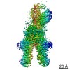
| Entry | Database: SASBDB / ID: SASDDF6 |
|---|---|
 Sample Sample | Myelin basic protein
|
| Function / homology |  Function and homology information Function and homology informationstructural constituent of myelin sheath / compact myelin / internode region of axon / myelination / cell periphery / phospholipid binding / myelin sheath / calmodulin binding / neuronal cell body / lipid binding ...structural constituent of myelin sheath / compact myelin / internode region of axon / myelination / cell periphery / phospholipid binding / myelin sheath / calmodulin binding / neuronal cell body / lipid binding / protein-containing complex / plasma membrane Similarity search - Function |
| Biological species |  |
 Citation Citation | Journal: J Am Chem Soc / Year: 2014 Title: Internal nanosecond dynamics in the intrinsically disordered myelin basic protein. Authors: Andreas M Stadler / Laura Stingaciu / Aurel Radulescu / Olaf Holderer / Michael Monkenbusch / Ralf Biehl / Dieter Richter /  Abstract: Intrinsically disordered proteins lack a well-defined folded structure and contain a high degree of structural freedom and conformational flexibility, which is expected to enhance binding to their ...Intrinsically disordered proteins lack a well-defined folded structure and contain a high degree of structural freedom and conformational flexibility, which is expected to enhance binding to their physiological targets. In solution and in the lipid-free state, myelin basic protein belongs to that class of proteins. Using small-angle scattering, the protein was found to be structurally disordered similar to Gaussian chains. The combination of structural and hydrodynamic information revealed an intermediary compactness of the protein between globular proteins and random coil polymers. Modeling by a coarse-grained structural ensemble gave indications for a compact core with flexible ends. Neutron spin-echo spectroscopy measurements revealed a large contribution of internal dynamics to the overall diffusion. The experimental results showed a high flexibility of the structural ensemble. Displacement patterns along the first two normal modes demonstrated that collective stretching and bending motions dominate the internal modes. The observed dynamics represent nanosecond conformational fluctuations within the reconstructed coarse-grained structural ensemble, allowing the exploration of a large configurational space. In an alternative approach, we investigated if models from polymer theory, recently used for the interpretation of fluorescence spectroscopy experiments on disordered proteins, are suitable for the interpretation of the observed motions. Within the framework of the Zimm model with internal friction (ZIF), a large offset of 81.6 ns is needed as an addition to all relaxation times due to intrachain friction sources. The ZIF model, however, shows small but systematic deviations from the measured data. The large value of the internal friction leads to the breakdown of the Zimm model. |
 Contact author Contact author |
|
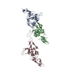
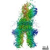
- Structure visualization
| Structure viewer | Molecule: MolmilJmol/JSmol |
|---|
- Downloads & links
-Data source
| SASBDB page |  SASDDF6 SASDDF6 |
|---|
-Related structure data
| Similar structure data |
|---|
-External links
| Related items in Molecule of the Month |
|---|

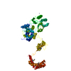
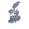
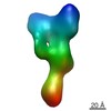
-Models
| Model #1987 |  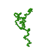 Type: mix / Radius of dummy atoms: 1.90 A / Chi-square value: 0.908209 / P-value: 0.000947  Search similar-shape structures of this assembly by Omokage search (details) Search similar-shape structures of this assembly by Omokage search (details) |
|---|---|
| Model #1988 | 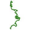 Type: mix / Radius of dummy atoms: 1.90 A / Chi-square value: 0.908209 / P-value: 0.000947 Search similar-shape structures of this assembly by Omokage search (details) |
| Model #1989 | 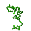 Type: mix / Radius of dummy atoms: 1.90 A / Chi-square value: 0.908209 / P-value: 0.000947 Search similar-shape structures of this assembly by Omokage search (details) |
| Model #1990 | 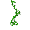 Type: mix / Radius of dummy atoms: 1.90 A / Chi-square value: 0.908209 / P-value: 0.000947 Search similar-shape structures of this assembly by Omokage search (details) |
| Model #1991 | 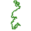 Type: mix / Radius of dummy atoms: 1.90 A / Chi-square value: 0.908209 / P-value: 0.000947 Search similar-shape structures of this assembly by Omokage search (details) |
| Model #1992 | 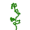 Type: mix / Radius of dummy atoms: 1.90 A / Chi-square value: 0.908209 / P-value: 0.000947 Search similar-shape structures of this assembly by Omokage search (details) |
-Sample
| Sample | Name: Myelin basic protein / Specimen concentration: 4.5 mg/ml |
|---|---|
| Buffer | Name: 20 mM NaH2PO4/ Na2HPO4, 99.9% D2O / pH: 4.8 / Comment: deuterium oxide |
| Entity #1062 | Type: protein / Description: Myelin basic protein / Formula weight: 18.323 / Num. of mol.: 1 / Source: Bos taurus / References: UniProt: P02687 Sequence: AAQKRPSQRS KYLASASTMD HARHGFLPRH RDTGILDSLG RFFGSDRGAP KRGSGKDGHH AARTTHYGSL PQKAQGHRPQ DENPVVHFFK NIVTPRTPPP SQGKGRGLSL SRFSWGAEGQ KPGFGYGGRA SDYKSAHKGL KGHDAQGTLS KIFKLGGRDS RSGSPMARR |
-Experimental information
| Beam | Instrument name: ESRF BM29 / City: Grenoble / 国: France  / Type of source: X-ray synchrotron / Wavelength: 0.099 Å / Type of source: X-ray synchrotron / Wavelength: 0.099 Å | |||||||||||||||||||||||||||||
|---|---|---|---|---|---|---|---|---|---|---|---|---|---|---|---|---|---|---|---|---|---|---|---|---|---|---|---|---|---|---|
| Detector | Name: Pilatus 1M / Type: Dectris / Pixsize x: 172 mm | |||||||||||||||||||||||||||||
| Scan | 
| |||||||||||||||||||||||||||||
| Distance distribution function P(R) | 
| |||||||||||||||||||||||||||||
| Result | 
|
