 Movie
Movie Controller
[English] 日本語
 Yorodumi
- SASDAX5: Endophilin-CoA complex (Endophilin-A1 BAR domain, Endophilin + ar... -
+ Open data

- Basic information
| Entry | Database: SASBDB / ID: SASDAX5 |
|---|---|
 Sample Sample | Endophilin-CoA complex
|
| Biological species |  |
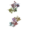
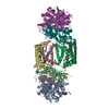
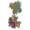
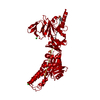
 Citation Citation | Journal: Front Mol Biosci / Year: 2014 Title: Endophilin-A1 BAR domain interaction with arachidonyl CoA. Authors: Maxim V Petoukhov / Winfried Weissenhorn / Dmitri I Svergun /   Abstract: Endophilin-A1 belongs to the family of BAR domain containing proteins that catalyze membrane remodeling processes via sensing, inducing and stabilizing membrane curvature. We show that the BAR domain ...Endophilin-A1 belongs to the family of BAR domain containing proteins that catalyze membrane remodeling processes via sensing, inducing and stabilizing membrane curvature. We show that the BAR domain of endophilin-A1 binds arachidonic acid and molds its coenzyme A (CoA) activated form, arachidonyl-CoA into a defined structure. We studied low resolution structures of endophilin-A1-BAR and its complex with arachidonyl-CoA in solution using synchrotron small-angle X-ray scattering (SAXS). The free endophilin-A1-BAR domain is shown to be dimeric at lower concentrations but builds tetramers and higher order complexes with increasing concentrations. Extensive titration SAXS studies revealed that the BAR domain produces a homogenous complex with the lipid micelles. The structural model of the complexes revealed two arachidonyl-CoA micelles bound to the distal arms of an endophilin-A1-BAR dimer. Intriguingly, the radius of the bound micelles significantly decreases compared to that of the free micelles, and this structural result may provide hints on the potential biological relevance of the endophilin-A1-BAR interaction with arachidonyl CoA. |
 Contact author Contact author |
|
- Structure visualization
| Structure viewer | Molecule:  MolmilJmol/JSmol MolmilJmol/JSmol |
|---|
- Downloads & links
 SASDAX5


-Models
| Model #143 |  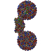 Type: dummy / Software: Sasref / Radius of dummy atoms: 1.90 A / Chi-square value: 24.01  Search similar-shape structures of this assembly by Omokage search (details) Search similar-shape structures of this assembly by Omokage search (details) |
|---|
-Sample
| Sample | Name: Endophilin-CoA complex / Sample MW: 118 kDa / Specimen concentration: 1 mg/ml / Entity id: 105 / 107 |
|---|---|
| Buffer | Name: TRIS-HCL / Concentration: 50.00 mM / pH: 8 / Composition: NaCl 300.000 mM |
| Entity #105 | Name: Endophilin / Type: protein / Description: Endophilin-A1 BAR domain / Formula weight: 29 / Num. of mol.: 2 / Source: Mus musculus Sequence: MSVAGLKKQF HKATQKVSEK VGGAEGTKLD DDFKEMERKV DVTSRAVMEI MTKTIEYLQP NPASRAKLSM INTMSKIRGQ EKGPGYPQAE ALLAEAMLKF GRELGDDCNF GPALGEVGEA MRELSEVKDS LDMEVKQNFI DPLQNLHDKD LREIQHHLKK LEGRRLDFDY ...Sequence: MSVAGLKKQF HKATQKVSEK VGGAEGTKLD DDFKEMERKV DVTSRAVMEI MTKTIEYLQP NPASRAKLSM INTMSKIRGQ EKGPGYPQAE ALLAEAMLKF GRELGDDCNF GPALGEVGEA MRELSEVKDS LDMEVKQNFI DPLQNLHDKD LREIQHHLKK LEGRRLDFDY KKKRQGKIPD EELRQALEKF DESKEIAESS MFNLLEMDIE QVSQLSALVQ AQLEYHKQAV QILQQVTVRL EERIRQA |
| Entity #107 | Name: CoA / Type: other / Description: arachidonyl-CoA |
-Experimental information
| Beam | Instrument name:  DORIS III X33 / City: Hamburg / 国: Germany / Type of source: X-ray synchrotron DORIS III X33 / City: Hamburg / 国: Germany / Type of source: X-ray synchrotron | |||||||||
|---|---|---|---|---|---|---|---|---|---|---|
| Detector | Name: MAR 345 Image Plate | |||||||||
| Scan | 
| |||||||||
| Result | I0 from Guinier: 435.06 / Rg from Guinier: 5.9 nm / D max: 19 / Type of curve: single_conc /
|
