Movie
Movie Controller


+ Open data

- Basic information
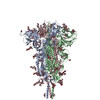
| Entry | Database: PDB / ID: 9zt5 | |||||||||||||||||||||||||||
|---|---|---|---|---|---|---|---|---|---|---|---|---|---|---|---|---|---|---|---|---|---|---|---|---|---|---|---|---|
| Title | SARS-CoV-2 S2 in complex with polyclonal Fab-B_Donor3 | |||||||||||||||||||||||||||
 Components Components |
| |||||||||||||||||||||||||||
 Keywords Keywords | VIRAL PROTEIN/Immune System / SARS-CoV-2 / Coronavirus / Immune system / antibody / VIRAL PROTEIN / VIRAL PROTEIN-Immune System complex | |||||||||||||||||||||||||||
| Function / homology |  Function and homology information Function and homology informationsymbiont-mediated disruption of host tissue / Maturation of spike protein / Translation of Structural Proteins / Virion Assembly and Release / host cell surface / host extracellular region / symbiont-mediated-mediated suppression of host tetherin activity / Induction of Cell-Cell Fusion / structural constituent of virion / positive regulation of viral entry into host cell ...symbiont-mediated disruption of host tissue / Maturation of spike protein / Translation of Structural Proteins / Virion Assembly and Release / host cell surface / host extracellular region / symbiont-mediated-mediated suppression of host tetherin activity / Induction of Cell-Cell Fusion / structural constituent of virion / positive regulation of viral entry into host cell / membrane fusion / host cell endoplasmic reticulum-Golgi intermediate compartment membrane / Attachment and Entry / entry receptor-mediated virion attachment to host cell / receptor-mediated virion attachment to host cell / host cell surface receptor binding / symbiont-mediated suppression of host innate immune response / endocytosis involved in viral entry into host cell / receptor ligand activity / fusion of virus membrane with host plasma membrane / fusion of virus membrane with host endosome membrane / viral envelope / symbiont entry into host cell / virion attachment to host cell / host cell plasma membrane / SARS-CoV-2 activates/modulates innate and adaptive immune responses / virion membrane / membrane / identical protein binding / plasma membrane Similarity search - Function | |||||||||||||||||||||||||||
| Biological species |   Severe acute respiratory syndrome coronavirus 2 Severe acute respiratory syndrome coronavirus 2 Homo sapiens (human) Homo sapiens (human) | |||||||||||||||||||||||||||
| Method | ELECTRON MICROSCOPY / single particle reconstruction / cryo EM / Resolution: 3.21 Å | |||||||||||||||||||||||||||
 Authors Authors | Park, S. / Ward, A.B. | |||||||||||||||||||||||||||
| Funding support |  United States, 1items United States, 1items
| |||||||||||||||||||||||||||
 Citation Citation | Journal: bioRxiv / Year: 2026 Title: The buried S2 apex of SARS-CoV-2 spike elicits an immunodominant germline-restricted public antibody response. Authors: Suncheol Park / Jacob Mischka / Nisreen Okba / Anass Abbad / Meng Yuan / Komal Srivastava / Charles Gleason / Lubbertus C F Mulder / Jeffrey Copps / Katrina Saam / Sandhya Bangaru / Florian ...Authors: Suncheol Park / Jacob Mischka / Nisreen Okba / Anass Abbad / Meng Yuan / Komal Srivastava / Charles Gleason / Lubbertus C F Mulder / Jeffrey Copps / Katrina Saam / Sandhya Bangaru / Florian Krammer / Ian A Wilson / Viviana Simon / Andrew B Ward /  Abstract: The continued mutational pressure on the SARS-CoV-2 S1 subunit underscores the need to target the conserved S2 region for pan-coronavirus vaccine development. A detailed molecular understanding of S2- ...The continued mutational pressure on the SARS-CoV-2 S1 subunit underscores the need to target the conserved S2 region for pan-coronavirus vaccine development. A detailed molecular understanding of S2-directed immune responses is therefore essential. In this study, we identified the S2 apex as the most immunodominant epitope within the S2 subunit, eliciting robust antibody responses despite occlusion by S1, using electron-microscopy-based polyclonal epitope mapping (EMPEM) of plasma from infected and vaccinated individuals. Structure-guided sequence analysis with antibody databases revealed that antibodies targeting a poorly characterized S2 Apex-B site form a convergent public clonotype, which is predominantly derived from the IGHV3-30 germline with a 14-residue CDRH3 containing a G/S-G-S/N-Y motif. This clonotype is extensively expanded, accounting for up to 40% of total spike-reactive antibody sequence counts in individual vaccinated donors. This study elucidates the molecular basis the high-frequency elicitation of this non-neutralizing clonotype emphasizing that its immunodominance acts as a primary hurdle for universal coronavirus vaccines and underscore the need for precision antigen design to redirect immunity toward more potent neutralizing targets. | |||||||||||||||||||||||||||
| History |
|
- Structure visualization
| Structure viewer | Molecule: MolmilJmol/JSmol |
|---|
- Downloads & links
-Download
| PDBx/mmCIF format | 9zt5.cif.gz | 336 KB | Display | PDBx/mmCIF format |
|---|---|---|---|---|
| PDB format | pdb9zt5.ent.gz | 218.2 KB | Display | PDB format |
| PDBx/mmJSON format | 9zt5.json.gz | Tree view | PDBx/mmJSON format | |
| Others |  Other downloads Other downloads |
-Validation report
| Arichive directory | https://data.pdbj.org/pub/pdb/validation_reports/zt/9zt5ftp://data.pdbj.org/pub/pdb/validation_reports/zt/9zt5 | HTTPS FTP |
|---|
-Related structure data
| Related structure data |  74737MC  10muC  9z80C  9zt6C  9zt7C  9zt8C M: map data used to model this data C: citing same article ( |
|---|---|
| Similar structure data |


-Links
 PDBj
PDBj


- Assembly
| Deposited unit | 
|
|---|---|
| 1 |
|
-Components
| #1: Antibody | Mass: 9670.944 Da / Num. of mol.: 1 / Source method: isolated from a natural source Details: This map was obtained from a polyclonal antibody in a serum sample. We built it as alanine. The cystein, consisting of a disulfide bond in a Fab, remains. Source: (natural) Homo sapiens (human) | ||||||||
|---|---|---|---|---|---|---|---|---|---|
| #2: Antibody | Mass: 8990.104 Da / Num. of mol.: 1 / Source method: isolated from a natural source Details: This map was obtained from a polyclonal antibody in a serum sample. We built it as alanine. The cystein, consisting of a disulfide bond in a Fab, remains. Source: (natural) Homo sapiens (human) | ||||||||
| #3: Protein | Mass: 52342.074 Da / Num. of mol.: 3 Source method: isolated from a genetically manipulated source Source: (gene. exp.) Severe acute respiratory syndrome coronavirus 2Gene: S, 2 / Production host: Homo sapiens (human) / References: UniProt: P0DTC2#4: Polysaccharide | 2-acetamido-2-deoxy-beta-D-glucopyranose-(1-4)-2-acetamido-2-deoxy-beta-D-glucopyranose Source method: isolated from a genetically manipulated source #5: Sugar | ChemComp-NAG /   Type: D-saccharide, beta linking / Mass: 221.208 Da / Num. of mol.: 9 / Source method: obtained synthetically / Formula: C8H15NO6 / Feature type: SUBJECT OF INVESTIGATION Type: D-saccharide, beta linking / Mass: 221.208 Da / Num. of mol.: 9 / Source method: obtained synthetically / Formula: C8H15NO6 / Feature type: SUBJECT OF INVESTIGATIONHas ligand of interest | Y | Has protein modification | Y | |
-Experimental details
-Experiment
| Experiment | Method: ELECTRON MICROSCOPY |
|---|---|
| EM experiment | Aggregation state: PARTICLE / 3D reconstruction method: single particle reconstruction |
- Sample preparation
| Component | Name: SARS-CoV-2 S2 in complex with polyclonal Fab-B_Donor3 / Type: COMPLEX / Entity ID: #1-#3 / Source: RECOMBINANT | |||||||||||||||
|---|---|---|---|---|---|---|---|---|---|---|---|---|---|---|---|---|
| Molecular weight | Experimental value: NO | |||||||||||||||
| Source (natural) | Organism: Homo sapiens (human) | |||||||||||||||
| Source (recombinant) | Organism: Homo sapiens (human) | |||||||||||||||
| Buffer solution | pH: 7.5 | |||||||||||||||
| Buffer component |
| |||||||||||||||
| Specimen | Conc.: 0.47 mg/ml / Embedding applied: NO / Shadowing applied: NO / Staining applied: NO / Vitrification applied: YES | |||||||||||||||
| Specimen support | Grid material: GOLD / Grid mesh size: 300 divisions/in. / Grid type: UltrAuFoil R1.2/1.3 | |||||||||||||||
| Vitrification | Instrument: FEI VITROBOT MARK IV / Cryogen name: ETHANE / Humidity: 100 % / Chamber temperature: 277 K |
- Electron microscopy imaging
| Microscopy | Model: TFS GLACIOS |
|---|---|
| Electron gun | Electron source:  FIELD EMISSION GUN / Accelerating voltage: 200 kV / Illumination mode: FLOOD BEAM FIELD EMISSION GUN / Accelerating voltage: 200 kV / Illumination mode: FLOOD BEAM |
| Electron lens | Mode: BRIGHT FIELD / Nominal magnification: 190000 X / Nominal defocus max: 1800 nm / Nominal defocus min: 800 nm / Cs: 2.7 mm / Alignment procedure: COMA FREE |
| Specimen holder | Cryogen: NITROGEN / Specimen holder model: FEI TITAN KRIOS AUTOGRID HOLDER |
| Image recording | Electron dose: 45 e/Å2 / Film or detector model: TFS FALCON 4i (4k x 4k) |
- Processing
| EM software |
| ||||||||||||||||||||||||||||||||||||
|---|---|---|---|---|---|---|---|---|---|---|---|---|---|---|---|---|---|---|---|---|---|---|---|---|---|---|---|---|---|---|---|---|---|---|---|---|---|
| CTF correction | Type: PHASE FLIPPING AND AMPLITUDE CORRECTION | ||||||||||||||||||||||||||||||||||||
| Symmetry | Point symmetry: C1 (asymmetric) | ||||||||||||||||||||||||||||||||||||
| 3D reconstruction | Resolution: 3.21 Å / Resolution method: FSC 0.143 CUT-OFF / Num. of particles: 38727 / Symmetry type: POINT | ||||||||||||||||||||||||||||||||||||
| Atomic model building | Space: REAL | ||||||||||||||||||||||||||||||||||||
| Atomic model building | PDB-ID: 6XR8 Accession code: 6XR8 / Source name: PDB / Type: experimental model | ||||||||||||||||||||||||||||||||||||
| Refinement | Cross valid method: NONE Stereochemistry target values: GeoStd + Monomer Library + CDL v1.2 | ||||||||||||||||||||||||||||||||||||
| Displacement parameters | Biso mean: 77.21 Å2 | ||||||||||||||||||||||||||||||||||||
| Refine LS restraints |
|