 Movie
Movie Controller

[English] 日本語
 Yorodumi
- PDB-9zfs: CryoEM structure of respiratory complex III intermediate from Aca... -
+ Open data

- Basic information
| Entry | Database: PDB / ID: 9zfs | |||||||||
|---|---|---|---|---|---|---|---|---|---|---|
| Title | CryoEM structure of respiratory complex III intermediate from Acanthamoeba castellanii | |||||||||
 Components Components |
| |||||||||
 Keywords Keywords | OXIDOREDUCTASE / complex III / OXPHOS / cytochrome bc1 complex | |||||||||
| Function / homology |  Function and homology information Function and homology informationpitrilysin / signal peptidase activity / respiratory chain complex III / quinol-cytochrome-c reductase activity / mitochondrial electron transport, ubiquinol to cytochrome c / metalloendopeptidase activity / mitochondrial inner membrane / mitochondrial matrix / metal ion binding / mitochondrion ...pitrilysin / signal peptidase activity / respiratory chain complex III / quinol-cytochrome-c reductase activity / mitochondrial electron transport, ubiquinol to cytochrome c / metalloendopeptidase activity / mitochondrial inner membrane / mitochondrial matrix / metal ion binding / mitochondrion / proteolysis / nucleus Similarity search - Function | |||||||||
| Biological species |  Acanthamoeba castellanii (eukaryote) Acanthamoeba castellanii (eukaryote) | |||||||||
| Method | ELECTRON MICROSCOPY / single particle reconstruction / cryo EM / Resolution: 3.65 Å | |||||||||
 Authors Authors | Fry, M.Y. / Luce, B.E. / Stefely, J.A. / Samuelson, J. / Mootha, V.K. / Chao, L.H. | |||||||||
| Funding support |  United States, 1items United States, 1items
| |||||||||
 Citation Citation | Journal: Cell / Year: 2026 Title: Acanthamoeba ATP synthase structure reveals the TCA cycle is tethered to OXPHOS. Authors: Michelle Y Fry / Bridget E Luce / Jonathan A Stefely / Michael Z Chen / Felicia G Deng / Tiana M Issa / / Sarah E Calvo / John Samuelson / Vamsi K Mootha / Luke H Chao / Abstract: Oxidative phosphorylation (OXPHOS) is a key metabolic process that couples redox energy to ATP production. While some core OXPHOS complex subunits are found across all domains of life, many have ...Oxidative phosphorylation (OXPHOS) is a key metabolic process that couples redox energy to ATP production. While some core OXPHOS complex subunits are found across all domains of life, many have diverged or expanded across evolution-as seen in the protozoan pathogen Acanthamoeba castellanii. By integrating cryo-electron microscopy of unenriched mitochondrial lysate with mass spectrometry proteomics, we resolved the structures of endogenous mitochondrial ATP synthase (complex V), Hsp60, and respiratory complex III from Acanthamoeba. We capture Acanthamoeba ATP synthase in an IF1-inhibited state and reveal how Acanthamoeba-specific subunits and extensions stabilize the molecular machine, which includes a β subunit extension that interfaces with the peripheral stalk. Additionally, we characterize an active malate dehydrogenase (MDH) dimer structurally integrated within the ATP synthase peripheral stalk, thus revealing a direct protein tether between OXPHOS and the tricarboxylic acid cycle. Together, these findings provide structural insight into lineage-specific adaptations in Acanthamoeba that may tune protozoan metabolism. | |||||||||
| History |
|
- Structure visualization
| Structure viewer | Molecule: MolmilJmol/JSmol |
|---|
- Downloads & links
-Download
| PDBx/mmCIF format | 9zfs.cif.gz | 1.1 MB | Display | PDBx/mmCIF format |
|---|---|---|---|---|
| PDB format | pdb9zfs.ent.gz | 898.7 KB | Display | PDB format |
| PDBx/mmJSON format | 9zfs.json.gz | Tree view | PDBx/mmJSON format | |
| Others |  Other downloads Other downloads |
-Validation report
| Arichive directory | https://data.pdbj.org/pub/pdb/validation_reports/zf/9zfsftp://data.pdbj.org/pub/pdb/validation_reports/zf/9zfs | HTTPS FTP |
|---|
-Related structure data
| Related structure data |  74150MC  9zfrC  9zftC  9zfuC  9zfvC  9zfwC  9zfxC M: map data used to model this data C: citing same article ( |
|---|---|
| Similar structure data |


-Links
 PDBj
PDBj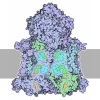
- Assembly
| Deposited unit | 
|
|---|---|
| 1 |
|
-Components
-Protein , 5 types, 10 molecules BCJHMIGRQT
| #1: Protein | Mass: 53933.297 Da / Num. of mol.: 2 / Source method: isolated from a natural source / Details: UQCRC2 | ACANB02868_t1 / Source: (natural) Acanthamoeba castellanii (eukaryote) / Strain: Neff / References: UniProt: L8GTW2#3: Protein | Mass: 54876.777 Da / Num. of mol.: 2 / Source method: isolated from a natural source / Details: PMPCB | ACANB00313_t1,PMPCB | ACANB00313_t1 / Source: (natural) Acanthamoeba castellanii (eukaryote) / Strain: NeffReferences: UniProt: L8HK40, mitochondrial processing peptidase #4: Protein | Mass: 14371.188 Da / Num. of mol.: 2 / Source method: isolated from a natural source / Details: UQCRB | ACANB09944 / Source: (natural) Acanthamoeba castellanii (eukaryote) / Strain: Neff / References: UniProt: L8H0Y7#7: Protein | Mass: 33652.250 Da / Num. of mol.: 2 / Source method: isolated from a natural source / Details: ACANB07833_t1 | CYC1 / Source: (natural) Acanthamoeba castellanii (eukaryote) / Strain: Neff#8: Protein | Mass: 43958.941 Da / Num. of mol.: 2 / Source method: isolated from a natural source / Details: MT CYB | ACANB15918 / Source: (natural) Acanthamoeba castellanii (eukaryote) / Strain: Neff / References: UniProt: Q37378 |
|---|
-Cytochrome b-c1 complex subunit ... , 4 types, 8 molecules EFOKANLW
| #2: Protein | Mass: 8151.524 Da / Num. of mol.: 2 / Source method: isolated from a natural source / Details: UQCRQ | ACANB04471 / Source: (natural) Acanthamoeba castellanii (eukaryote) / Strain: Neff / References: UniProt: L8GXE4#5: Protein | Mass: 9282.360 Da / Num. of mol.: 2 / Source method: isolated from a natural source / Details: UQCR10 | ACANB07539 / Source: (natural) Acanthamoeba castellanii (eukaryote) / Strain: Neff / References: UniProt: L8HKA7#6: Protein | Mass: 8013.151 Da / Num. of mol.: 2 / Source method: isolated from a natural source / Details: UQCRH| ACANB09203_t1 / Source: (natural) Acanthamoeba castellanii (eukaryote) / Strain: Neff / References: UniProt: L8H1V3#9: Protein | Mass: 10705.308 Da / Num. of mol.: 2 / Source method: isolated from a natural source / Details: UQCR11 | ACANB09854_t1 / Source: (natural) Acanthamoeba castellanii (eukaryote) / Strain: Neff / References: UniProt: L8HFA4 |
|---|
-Non-polymers , 1 types, 4 molecules 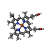
| #10: Chemical | ChemComp-HEM / 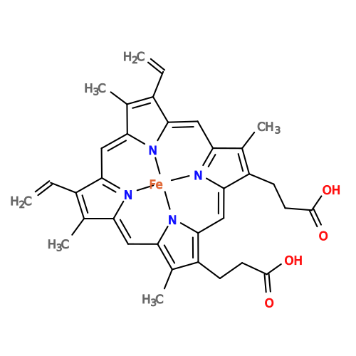 Mass: 616.487 Da / Num. of mol.: 4 / Source method: obtained synthetically / Formula: C34H32FeN4O4 / Feature type: SUBJECT OF INVESTIGATION Mass: 616.487 Da / Num. of mol.: 4 / Source method: obtained synthetically / Formula: C34H32FeN4O4 / Feature type: SUBJECT OF INVESTIGATION |
|---|
-Details
| Has ligand of interest | Y |
|---|---|
| Has protein modification | Y |
-Experimental details
-Experiment
| Experiment | Method: ELECTRON MICROSCOPY |
|---|---|
| EM experiment | Aggregation state: PARTICLE / 3D reconstruction method: single particle reconstruction |
- Sample preparation
| Component | Name: Acanthamoeba castellanii OXPHOS complex III intermediate assembly state missing the Rieske subunit Type: COMPLEX / Entity ID: #1-#9 / Source: NATURAL | ||||||||||||||||||||||||||||
|---|---|---|---|---|---|---|---|---|---|---|---|---|---|---|---|---|---|---|---|---|---|---|---|---|---|---|---|---|---|
| Molecular weight | Value: 1.3 MDa / Experimental value: NO | ||||||||||||||||||||||||||||
| Source (natural) | Organism: Acanthamoeba castellanii (eukaryote) / Strain: Neff | ||||||||||||||||||||||||||||
| Buffer solution | pH: 7.9 | ||||||||||||||||||||||||||||
| Buffer component |
| ||||||||||||||||||||||||||||
| Specimen | Conc.: 2.25 mg/ml / Embedding applied: NO / Shadowing applied: NO / Staining applied: NO / Vitrification applied: YES Details: Sample was not purified. Instead it was solved from imaging mitochondrial lysate. | ||||||||||||||||||||||||||||
| Vitrification | Instrument: FEI VITROBOT MARK IV / Cryogen name: ETHANE-PROPANE / Humidity: 100 % / Chamber temperature: 277.14 K |
- Electron microscopy imaging
| Experimental equipment |  Model: Titan Krios / Image courtesy: FEI Company |
|---|---|
| Microscopy | Model: TFS KRIOS |
| Electron gun | Electron source:  FIELD EMISSION GUN / Accelerating voltage: 300 kV / Illumination mode: SPOT SCAN FIELD EMISSION GUN / Accelerating voltage: 300 kV / Illumination mode: SPOT SCAN |
| Electron lens | Mode: DARK FIELD / Nominal magnification: 105000 X / Nominal defocus max: 2000 nm / Nominal defocus min: 600 nm |
| Specimen holder | Cryogen: NITROGEN / Specimen holder model: FEI TITAN KRIOS AUTOGRID HOLDER |
| Image recording | Electron dose: 0.99 e/Å2 / Film or detector model: GATAN K3 BIOQUANTUM (6k x 4k) |
| EM imaging optics | Energyfilter name: GIF Bioquantum / Energyfilter slit width: 20 eV |
- Processing
| EM software |
| |||||||||||||||||||||||||||||||||||
|---|---|---|---|---|---|---|---|---|---|---|---|---|---|---|---|---|---|---|---|---|---|---|---|---|---|---|---|---|---|---|---|---|---|---|---|---|
| CTF correction | Type: NONE | |||||||||||||||||||||||||||||||||||
| 3D reconstruction | Resolution: 3.65 Å / Resolution method: FSC 0.143 CUT-OFF / Num. of particles: 5208 / Symmetry type: POINT | |||||||||||||||||||||||||||||||||||
| Refinement | Highest resolution: 3.65 Å Stereochemistry target values: REAL-SPACE (WEIGHTED MAP SUM AT ATOM CENTERS) | |||||||||||||||||||||||||||||||||||
| Refine LS restraints |
|
