 Movie
Movie Controller

[English] 日本語
 Yorodumi
- EMDB-74156: Membrane region of Acanthamoeba castellanii mitochondrial ATP syn... -
+ Open data

- Basic information
| Entry |  | |||||||||
|---|---|---|---|---|---|---|---|---|---|---|
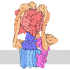
| Title | Membrane region of Acanthamoeba castellanii mitochondrial ATP synthase dimer | |||||||||
 Map data Map data | ||||||||||
 Sample Sample |
| |||||||||
 Keywords Keywords | complex V / ATP synthase / OXPHOS complex V / HYDROLASE | |||||||||
| Function / homology |  Function and homology information Function and homology informationproton-transporting ATP synthase complex / proton-transporting ATP synthase activity, rotational mechanism / mitochondrial membrane / mitochondrial inner membrane Similarity search - Function | |||||||||
| Biological species |  Acanthamoeba castellanii (eukaryote) Acanthamoeba castellanii (eukaryote) | |||||||||
| Method | single particle reconstruction / cryo EM / Resolution: 4.23 Å | |||||||||
 Authors Authors | Fry MY / Luce BE / Stefely JA / Mootha VK / Chao LH | |||||||||
| Funding support |  United States, 1 items United States, 1 items
| |||||||||
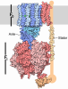
 Citation Citation | Journal: Cell / Year: 2026 Title: Acanthamoeba ATP synthase structure reveals the TCA cycle is tethered to OXPHOS. Authors: Michelle Y Fry / Bridget E Luce / Jonathan A Stefely / Michael Z Chen / Felicia G Deng / Tiana M Issa / / Sarah E Calvo / John Samuelson / Vamsi K Mootha / Luke H Chao / Abstract: Oxidative phosphorylation (OXPHOS) is a key metabolic process that couples redox energy to ATP production. While some core OXPHOS complex subunits are found across all domains of life, many have ...Oxidative phosphorylation (OXPHOS) is a key metabolic process that couples redox energy to ATP production. While some core OXPHOS complex subunits are found across all domains of life, many have diverged or expanded across evolution-as seen in the protozoan pathogen Acanthamoeba castellanii. By integrating cryo-electron microscopy of unenriched mitochondrial lysate with mass spectrometry proteomics, we resolved the structures of endogenous mitochondrial ATP synthase (complex V), Hsp60, and respiratory complex III from Acanthamoeba. We capture Acanthamoeba ATP synthase in an IF1-inhibited state and reveal how Acanthamoeba-specific subunits and extensions stabilize the molecular machine, which includes a β subunit extension that interfaces with the peripheral stalk. Additionally, we characterize an active malate dehydrogenase (MDH) dimer structurally integrated within the ATP synthase peripheral stalk, thus revealing a direct protein tether between OXPHOS and the tricarboxylic acid cycle. Together, these findings provide structural insight into lineage-specific adaptations in Acanthamoeba that may tune protozoan metabolism. | |||||||||
| History |
|
- Structure visualization
| Supplemental images |
|---|

- Downloads & links
-EMDB archive
| Map data | emd_74156.map.gz | 229.6 MB | EMDB map data format | |
|---|---|---|---|---|
| Header (meta data) | emd-74156-v30.xmlemd-74156.xml | 24.9 KB 24.9 KB | Display Display | EMDB header |
| Images |  emd_74156.png emd_74156.png | 42.4 KB | ||
| Filedesc metadata | emd-74156.cif.gz | 6.7 KB | ||
| Others | emd_74156_half_map_1.map.gzemd_74156_half_map_2.map.gz | 226.1 MB 226.1 MB | ||
| Archive directory |  https://data.pdbj.org/pub/emdb/structures/EMD-74156ftp://data.pdbj.org/pub/emdb/structures/EMD-74156 https://data.pdbj.org/pub/emdb/structures/EMD-74156ftp://data.pdbj.org/pub/emdb/structures/EMD-74156 | HTTPS FTP |
-Related structure data
| Related structure data |  9zfwMC  9zfrC  9zfsC  9zftC  9zfuC  9zfvC  9zfxC M: atomic model generated by this map C: citing same article ( |
|---|---|
| Similar structure data |


-Links
| EMDB pages | EMDB (EBI/PDBe) / EMDataResource |
|---|---|
| Related items in Molecule of the Month |
-Map
| File | Download / File: emd_74156.map.gz / Format: CCP4 / Size: 244.1 MB / Type: IMAGE STORED AS FLOATING POINT NUMBER (4 BYTES) | ||||||||||||||||||||||||||||||||||||
|---|---|---|---|---|---|---|---|---|---|---|---|---|---|---|---|---|---|---|---|---|---|---|---|---|---|---|---|---|---|---|---|---|---|---|---|---|---|
| Projections & slices | Image control
Images are generated by Spider. | ||||||||||||||||||||||||||||||||||||
| Voxel size | X=Y=Z: 0.822 Å | ||||||||||||||||||||||||||||||||||||
| Density |
| ||||||||||||||||||||||||||||||||||||
| Symmetry | Space group: 1 | ||||||||||||||||||||||||||||||||||||
| Details | EMDB XML:
|
 Z (Sec.)
 Y (Row.)
 X (Col.)


-Supplemental data
-Half map: #1
| File | emd_74156_half_map_1.map | ||||||||||||
|---|---|---|---|---|---|---|---|---|---|---|---|---|---|
| Projections & Slices |
| ||||||||||||
| Density Histograms |


-Half map: #2
| File | emd_74156_half_map_2.map | ||||||||||||
|---|---|---|---|---|---|---|---|---|---|---|---|---|---|
| Projections & Slices |
| ||||||||||||
| Density Histograms |


- Sample components
-Entire : Acanthamoeba castellanii OXPHOS complex III intermediate assembly...
| Entire | Name: Acanthamoeba castellanii OXPHOS complex III intermediate assembly state missing the Rieske subunit |
|---|---|
| Components |
|
-Supramolecule #1: Acanthamoeba castellanii OXPHOS complex III intermediate assembly...
| Supramolecule | Name: Acanthamoeba castellanii OXPHOS complex III intermediate assembly state missing the Rieske subunit type: complex / ID: 1 / Parent: 0 / Macromolecule list: #1-#5, #7, #6 |
|---|---|
| Source (natural) | Organism: Acanthamoeba castellanii (eukaryote) / Strain: Neff |
| Molecular weight | Theoretical: 1.3 MDa |
-Macromolecule #1: ATP synthase subunit e, mitochondrial
| Macromolecule | Name: ATP synthase subunit e, mitochondrial / type: protein_or_peptide / ID: 1 / Details: ACANB06593|subunit e / Number of copies: 2 / Enantiomer: LEVO |
|---|---|
| Source (natural) | Organism: Acanthamoeba castellanii (eukaryote) / Strain: Neff |
| Molecular weight | Theoretical: 11.392696 KDa |
| Sequence | String: MATPLQVPCG CRNSLQSFGA AAAFTVAFVW TSQSVQVKRN AYFSNLKSEI DTLKKKNHDL EHEVELLKNP PEPKEAHTDF AADFESGNI DFEKILEQVA SDH UniProtKB: Uncharacterized protein |
-Macromolecule #2: ATP synthase subunit g, mitochondrial
| Macromolecule | Name: ATP synthase subunit g, mitochondrial / type: protein_or_peptide / ID: 2 / Details: ACANB02343 | subunit g / Number of copies: 2 / Enantiomer: LEVO |
|---|---|
| Source (natural) | Organism: Acanthamoeba castellanii (eukaryote) / Strain: Neff |
| Molecular weight | Theoretical: 12.001693 KDa |
| Sequence | String: MNRLCGSLAK GAQKFQALEQ STTSCACNKQ LYGYLGTQFG CWSNRWQAFP ADAKALWRRV ITGQITVGEV GRGVRASTPI LLAALAGNF LGRGEFLYSP IVGASYTYPE Y |
-Macromolecule #3: ATP synthase subunit d, mitochondrial
| Macromolecule | Name: ATP synthase subunit d, mitochondrial / type: protein_or_peptide / ID: 3 / Details: ACANB10705|subunit d / Number of copies: 2 / Enantiomer: LEVO |
|---|---|
| Source (natural) | Organism: Acanthamoeba castellanii (eukaryote) / Strain: Neff |
| Molecular weight | Theoretical: 56.230902 KDa |
| Sequence | String: MRRISRVVAS SARGTALNKS SRTAAVFSRG YAAPETPQTD FKVTDAGIRV YAHGVVHDMN FLGDLPADAQ EVLSGKEWVP QSVSDFTAR WEKDAPELAN QARQLESLIT KFEKDPVQFG KSEAYLRDIA IHTTLERAAA RDVSPVEIAL THFWTAYSEV P SGGKLRES ...String: MRRISRVVAS SARGTALNKS SRTAAVFSRG YAAPETPQTD FKVTDAGIRV YAHGVVHDMN FLGDLPADAQ EVLSGKEWVP QSVSDFTAR WEKDAPELAN QARQLESLIT KFEKDPVQFG KSEAYLRDIA IHTTLERAAA RDVSPVEIAL THFWTAYSEV P SGGKLRES APTQLERVKA FLQSKKDPEL SAQNSRARKT AAIVKFVQAS SAEDKAAAAA LLLGSEKGQA LKAALKGVND AA KKEQIVK QHLQWDEATQ AAAELTQRHP EFTELKEVLA HFPPLVHHIG EPSAVQYRFK HATDKAYKKW QQNYSLTSSD EVT EKDPLL KNLRELLYSG KPANTVADAK KAQALQRSFP GLLFSRDVES FSNFVRKDEE VKSQLSTIYE DSLKEYPNDP ALAV PKDLD QRVKLWSKHT QELADVQQQW LDAKIAFIDA KIQEGMVQMD KFVNGTVDDI LADHPEWEKE IEDDIANHRW DPEME KKEY DNALHHYYEH HTVHA UniProtKB: Uncharacterized protein |
-Macromolecule #4: ATP synthase subunit 8, mitochondrial
| Macromolecule | Name: ATP synthase subunit 8, mitochondrial / type: protein_or_peptide / ID: 4 / Details: ACANB15938|subunit 8 / Number of copies: 2 / Enantiomer: LEVO |
|---|---|
| Source (natural) | Organism: Acanthamoeba castellanii (eukaryote) / Strain: Neff |
| Molecular weight | Theoretical: 17.42434 KDa |
| Sequence | String: MPQLDKLSFA TQYFWLTLFF FGLYFLSVNF FVILVFKNLK LRNIIYKIWY FFLYRFDYVD YNNKHKSLVN TSFSFIYYTL YINFFILTK INFIFEKTKS IINKTQTLNQ SQRLLMISLL NNFLNSNSVD FVKKFQELNI DEI UniProtKB: ORFB |
-Macromolecule #5: ATP synthase subunit b, mitochondrial
| Macromolecule | Name: ATP synthase subunit b, mitochondrial / type: protein_or_peptide / ID: 5 / Details: >ACANB15939|subunit b / Number of copies: 2 / Enantiomer: LEVO |
|---|---|
| Source (natural) | Organism: Acanthamoeba castellanii (eukaryote) / Strain: Neff |
| Molecular weight | Theoretical: 14.401209 KDa |
| Sequence | String: MKYSILLFFT LFAINNLLHY ISEKVIIVIF IVFLFFLMGF LSNIINSEFK SKIDYILNDI NAYVNVVFNF LYINKLFYLN LKSLKTGFL SLNSILGKKI DSLKFYTTLS NKLNAISSTT VLLLK UniProtKB: ORF25 |
-Macromolecule #6: ATP synthase subunit a
| Macromolecule | Name: ATP synthase subunit a / type: protein_or_peptide / ID: 6 / Details: ACANB15943|ATP6 / Number of copies: 2 / Enantiomer: LEVO |
|---|---|
| Source (natural) | Organism: Acanthamoeba castellanii (eukaryote) / Strain: Neff |
| Molecular weight | Theoretical: 28.654133 KDa |
| Sequence | String: MIFNPLEQFR ISVLQKLFFG NIDISITNNT IILFVILIGF TFLFYVNYST NTYIPSKWQY AVENIYLFVL QLFKQQINNI VALKYFPLV LFVFSFILFA NLIGLLPYGF TITGHIIFTF QIAFSLFFGI TLINFFNNKT EFFNLFVPSG VPKPLIPFLV V IEVVSYLI ...String: MIFNPLEQFR ISVLQKLFFG NIDISITNNT IILFVILIGF TFLFYVNYST NTYIPSKWQY AVENIYLFVL QLFKQQINNI VALKYFPLV LFVFSFILFA NLIGLLPYGF TITGHIIFTF QIAFSLFFGI TLINFFNNKT EFFNLFVPSG VPKPLIPFLV V IEVVSYLI RPFSLSVRLF ANMLAGHTLL NILSAFIFNV FKKYALISFL PLLFIVFIIV LEFCIAIVQA YIFSILTCIY LN DIYNTSH UniProtKB: ATP synthase subunit a |
-Macromolecule #7: ATP synthase subunit f, mitochondrial
| Macromolecule | Name: ATP synthase subunit f, mitochondrial / type: protein_or_peptide / ID: 7 / Details: ACANB14194|subunit f / Number of copies: 2 / Enantiomer: LEVO |
|---|---|
| Source (natural) | Organism: Acanthamoeba castellanii (eukaryote) / Strain: Neff |
| Molecular weight | Theoretical: 10.670317 KDa |
| Sequence | String: MSRSLITRSL PLRSSSHGAL GGARPVFSEV PLKEVPSYVR LHVLGNTRRL TKEFVQWYWR TYFHAGKADP IIHGILLVSG VGYLLHNHW HGSHP UniProtKB: Uncharacterized protein |
-Macromolecule #8: ATP synthase subunit i/j, mitochondrial
| Macromolecule | Name: ATP synthase subunit i/j, mitochondrial / type: protein_or_peptide / ID: 8 / Details: ACANB04724|subunit i/j / Number of copies: 2 / Enantiomer: LEVO |
|---|---|
| Source (natural) | Organism: Acanthamoeba castellanii (eukaryote) / Strain: Neff |
| Molecular weight | Theoretical: 10.638169 KDa |
| Sequence | String: MSARNFGLTE GEIQELKKRP YPRFAPAPMQ RLISQAKVIW PFAVGWAVTL FLYSRIPISD DDRAKSHYQY QLDVLDGKIK PEDHPIHGH HGH |
-Experimental details
-Structure determination
| Method | cryo EM |
|---|---|
 Processing Processing | single particle reconstruction |
| Aggregation state | particle |
-Sample preparation
| Concentration | 2.25 mg/mL | ||||||||||||||
|---|---|---|---|---|---|---|---|---|---|---|---|---|---|---|---|
| Buffer | pH: 7.9 Component:
| ||||||||||||||
| Vitrification | Cryogen name: ETHANE-PROPANE / Chamber humidity: 100 % / Chamber temperature: 277.14 K / Instrument: FEI VITROBOT MARK IV | ||||||||||||||
| Details | Sample was not purified. Instead it was solved from imaging mitochondrial lysate. |
- Electron microscopy
| Microscope | TFS KRIOS |
|---|---|
| Specialist optics | Energy filter - Name: GIF Bioquantum / Energy filter - Slit width: 20 eV |
| Image recording | Film or detector model: GATAN K3 BIOQUANTUM (6k x 4k) / Average electron dose: 0.99 e/Å2 |
| Electron beam | Acceleration voltage: 300 kV / Electron source:  FIELD EMISSION GUN FIELD EMISSION GUN |
| Electron optics | Illumination mode: SPOT SCAN / Imaging mode: DARK FIELD / Nominal defocus max: 2.0 µm / Nominal defocus min: 0.6 µm / Nominal magnification: 105000 |
| Sample stage | Specimen holder model: FEI TITAN KRIOS AUTOGRID HOLDER / Cooling holder cryogen: NITROGEN |
| Experimental equipment |  Model: Titan Krios / Image courtesy: FEI Company |
