 Movie
Movie Controller


+ Open data

- Basic information
| Entry | Database: PDB / ID: 9ygl | |||||||||
|---|---|---|---|---|---|---|---|---|---|---|
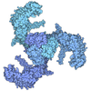
| Title | RCK Gating Ring from Kch in the closed conformation | |||||||||
 Components Components | (Voltage-gated potassium channel Kch) x 2 | |||||||||
 Keywords Keywords | TRANSPORT PROTEIN / Ion Channel Gating ring / RCK / Potassium Channel / MEMBRANE PROTEIN | |||||||||
| Function / homology |  Function and homology information Function and homology informationpotassium channel activity / potassium ion transport / potassium ion transmembrane transport / protein-containing complex / identical protein binding / plasma membrane Similarity search - Function | |||||||||
| Biological species |   Escherichia coli (E. coli) Escherichia coli (E. coli) | |||||||||
| Method | ELECTRON MICROSCOPY / single particle reconstruction / cryo EM / Resolution: 2.88 Å | |||||||||
 Authors Authors | Morote-Costas, B. / Zhou, M. | |||||||||
| Funding support | 1items
| |||||||||
 Citation Citation | Journal: PNAS Nexus / Year: 2026 Title: Mechanism of Kch activation by Zn. Authors: Brian Morote-Costas / Yaping Pan / Lie Wang / Xiaochen Bai / Steve W Lockless / Ming Zhou /  Abstract: has a single K channel gene, , that produces a K channel with six transmembrane segments and a cytosolic Regulate Conductance of K channels (RCK) domain. RCK domains are present in both eukaryotic ... has a single K channel gene, , that produces a K channel with six transmembrane segments and a cytosolic Regulate Conductance of K channels (RCK) domain. RCK domains are present in both eukaryotic and prokaryotic organisms and known to regulate ion channel activity upon binding of an ion or a nucleotide, however, how Kch is regulated remains unknown. Here, we show that Zn or Cu activates Kch via the RCK domain. Structural studies reveal that eight RCK domains assemble into a gating ring that can assume three conformations, closed, intermediate, and open, and that Zn promotes the open conformation by stabilizing the assembly interface. These results expand our knowledge on Kch and on RCK-mediated regulation of ion channels. | |||||||||
| History |
|
- Structure visualization
| Structure viewer | Molecule: MolmilJmol/JSmol |
|---|
- Downloads & links
-Download
| PDBx/mmCIF format | 9ygl.cif.gz | 248.8 KB | Display | PDBx/mmCIF format |
|---|---|---|---|---|
| PDB format | pdb9ygl.ent.gz | 192.1 KB | Display | PDB format |
| PDBx/mmJSON format | 9ygl.json.gz | Tree view | PDBx/mmJSON format | |
| Others |  Other downloads Other downloads |
-Validation report
| Arichive directory | https://data.pdbj.org/pub/pdb/validation_reports/yg/9yglftp://data.pdbj.org/pub/pdb/validation_reports/yg/9ygl | HTTPS FTP |
|---|
-Related structure data
| Related structure data |  72932MC  9ye0C  9yfvC  9ykqC C: citing same article ( M: map data used to model this data |
|---|---|
| Similar structure data |


-Links
 PDBj
PDBj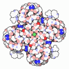
- Assembly
| Deposited unit | 
|
|---|---|
| 1 |
|
-Components
| #1: Protein | Mass: 19456.959 Da / Num. of mol.: 4 Source method: isolated from a genetically manipulated source Details: shorter isoform, beginning with alternate start codon at M240 Source: (gene. exp.) Escherichia coli (E. coli) / Strain: K-12 / Gene: kch, b1250, JW1242 / Production host: Escherichia coli BL21(DE3) (bacteria) / References: UniProt: P31069#2: Protein | Mass: 46105.371 Da / Num. of mol.: 4 Source method: isolated from a genetically manipulated source Source: (gene. exp.) Escherichia coli (E. coli) / Strain: K-12 / Gene: kch, b1250, JW1242 / Production host: Escherichia coli BL21(DE3) (bacteria) / References: UniProt: P31069Has protein modification | N | |
|---|
-Experimental details
-Experiment
| Experiment | Method: ELECTRON MICROSCOPY |
|---|---|
| EM experiment | Aggregation state: PARTICLE / 3D reconstruction method: single particle reconstruction |
- Sample preparation
| Component | Name: RCK Domain Dimer / Type: COMPLEX / Entity ID: #1 / Source: RECOMBINANT | ||||||||||||||||||||
|---|---|---|---|---|---|---|---|---|---|---|---|---|---|---|---|---|---|---|---|---|---|
| Molecular weight | Value: 261.914 MDa / Experimental value: YES | ||||||||||||||||||||
| Source (natural) | Organism: Escherichia coli (E. coli) / Strain: K-12 | ||||||||||||||||||||
| Source (recombinant) | Organism: Escherichia coli BL21(DE3) (bacteria) | ||||||||||||||||||||
| Buffer solution | pH: 7.4 | ||||||||||||||||||||
| Buffer component |
| ||||||||||||||||||||
| Specimen | Conc.: 8 mg/ml / Embedding applied: NO / Shadowing applied: NO / Staining applied: NO / Vitrification applied: YES | ||||||||||||||||||||
| Vitrification | Instrument: FEI VITROBOT MARK IV / Cryogen name: ETHANE / Humidity: 70 % / Chamber temperature: 281.15 K |
- Electron microscopy imaging
| Experimental equipment |  Model: Titan Krios / Image courtesy: FEI Company |
|---|---|
| Microscopy | Model: TFS KRIOS |
| Electron gun | Electron source:  FIELD EMISSION GUN / Accelerating voltage: 300 kV / Illumination mode: SPOT SCAN FIELD EMISSION GUN / Accelerating voltage: 300 kV / Illumination mode: SPOT SCAN |
| Electron lens | Mode: BRIGHT FIELD / Nominal magnification: 81000 X / Nominal defocus max: 800 nm / Nominal defocus min: 200 nm / Cs: 2.7 mm |
| Specimen holder | Cryogen: NITROGEN / Specimen holder model: FEI TITAN KRIOS AUTOGRID HOLDER |
| Image recording | Electron dose: 50 e/Å2 / Film or detector model: GATAN K3 (6k x 4k) |
- Processing
| EM software |
| ||||||||||||||||||||||||
|---|---|---|---|---|---|---|---|---|---|---|---|---|---|---|---|---|---|---|---|---|---|---|---|---|---|
| CTF correction | Type: PHASE FLIPPING AND AMPLITUDE CORRECTION | ||||||||||||||||||||||||
| Particle selection | Num. of particles selected: 18170630 | ||||||||||||||||||||||||
| 3D reconstruction | Resolution: 2.88 Å / Resolution method: FSC 0.143 CUT-OFF / Num. of particles: 55071 / Symmetry type: POINT | ||||||||||||||||||||||||
| Refinement | Highest resolution: 2.88 Å Stereochemistry target values: REAL-SPACE (WEIGHTED MAP SUM AT ATOM CENTERS) | ||||||||||||||||||||||||
| Refine LS restraints |
|
