 Movie
Movie Controller

[English] 日本語
 Yorodumi
- PDB-9y6o: Structure of fimbriae-like lipoprotein by Cryo Electron Microscopy -
+ Open data

- Basic information
| Entry | Database: PDB / ID: 9y6o | |||||||||
|---|---|---|---|---|---|---|---|---|---|---|
| Title | Structure of fimbriae-like lipoprotein by Cryo Electron Microscopy | |||||||||
 Components Components | Minor fimbrium subunit Mfa1 C-terminal domain-containing protein | |||||||||
 Keywords Keywords | CELL ADHESION / ffp1 / porphyromonas gingivalis / pili | |||||||||
| Function / homology | Fimbrial subunit protein, C-terminal / Major fimbrial subunit protein type IV, Fimbrillin, C-terminal / Prokaryotic membrane lipoprotein lipid attachment site profile. / Minor fimbrium subunit Mfa1 C-terminal domain-containing protein Function and homology information Function and homology information | |||||||||
| Biological species |  Porphyromonas gingivalis W83 (bacteria) Porphyromonas gingivalis W83 (bacteria) | |||||||||
| Method | ELECTRON MICROSCOPY / helical reconstruction / cryo EM / Resolution: 3.2 Å | |||||||||
 Authors Authors | Hanssen, E. / Gorasia, D.G. / Reynolds, E.C. | |||||||||
| Funding support |  Australia, 1items Australia, 1items
| |||||||||
 Citation Citation | Journal: Res Sq / Year: 2026 Title: Novel quadruple helical assembly of a Type V pilin in Porphyromonas gingivalis Authors: Reynolds, E.C. / Gorasia, D.G. / Slakeski, N. / Gui, M. / Chen, Y.-Y. / Moore, C. / Catmull, D. / Veith, P. / Dashper, S. / Hanssen, E. | |||||||||
| History |
|
- Structure visualization
| Structure viewer | Molecule: MolmilJmol/JSmol |
|---|
- Downloads & links
-Download
| PDBx/mmCIF format | 9y6o.cif.gz | 159.8 KB | Display | PDBx/mmCIF format |
|---|---|---|---|---|
| PDB format | pdb9y6o.ent.gz | 123.9 KB | Display | PDB format |
| PDBx/mmJSON format | 9y6o.json.gz | Tree view | PDBx/mmJSON format | |
| Others |  Other downloads Other downloads |
-Validation report
| Arichive directory | https://data.pdbj.org/pub/pdb/validation_reports/y6/9y6oftp://data.pdbj.org/pub/pdb/validation_reports/y6/9y6o | HTTPS FTP |
|---|
-Related structure data
| Related structure data |  72625MC M: map data used to model this data C: citing same article ( |
|---|---|
| Similar structure data |
-Links
 PDBj
PDBj
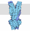
- Assembly
| Deposited unit | 
|
|---|---|
| 1 | x 12
|
| 2 |
|
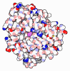
| Symmetry | Helical symmetry: (Circular symmetry: 2 / Dyad axis: no / N subunits divisor: 1 / Num. of operations: 6 / Rise per n subunits: 60.43 Å / Rotation per n subunits: 25.86 °) |
-Components
| #1: Protein | Mass: 46953.746 Da / Num. of mol.: 2 / Source method: isolated from a natural source / Source: (natural) Porphyromonas gingivalis W83 (bacteria) / References: UniProt: Q7MTR3#2: Polysaccharide | Type: oligosaccharide / Mass: 796.719 Da / Num. of mol.: 2 / Source method: isolated from a natural source Has ligand of interest | N | Has protein modification | Y | |
|---|
-Experimental details
-Experiment
| Experiment | Method: ELECTRON MICROSCOPY |
|---|---|
| EM experiment | Aggregation state: FILAMENT / 3D reconstruction method: helical reconstruction |
- Sample preparation
| Component | Name: ffp1 a type V pilin / Type: COMPLEX / Entity ID: #1 / Source: NATURAL | |||||||||||||||||||||||||
|---|---|---|---|---|---|---|---|---|---|---|---|---|---|---|---|---|---|---|---|---|---|---|---|---|---|---|
| Molecular weight | Experimental value: NO | |||||||||||||||||||||||||
| Source (natural) | Organism: Porphyromonas gingivalis (bacteria) / Strain: W83 | |||||||||||||||||||||||||
| Buffer solution | pH: 7.5 | |||||||||||||||||||||||||
| Buffer component |
| |||||||||||||||||||||||||
| Specimen | Embedding applied: NO / Shadowing applied: NO / Staining applied: NO / Vitrification applied: YES | |||||||||||||||||||||||||
| Specimen support | Details: 15 mA / Grid material: COPPER / Grid type: Quantifoil R1.2/1.3 | |||||||||||||||||||||||||
| Vitrification | Instrument: FEI VITROBOT MARK IV / Cryogen name: ETHANE / Humidity: 95 % / Chamber temperature: 295 K Details: Blot Force -1 Blot time 3 sec temp 22 degres C sample size 4ul |
- Electron microscopy imaging
| Experimental equipment |  Model: Titan Krios / Image courtesy: FEI Company |
|---|---|
| Microscopy | Model: TFS KRIOS |
| Electron gun | Electron source:  FIELD EMISSION GUN / Accelerating voltage: 300 kV / Illumination mode: FLOOD BEAM FIELD EMISSION GUN / Accelerating voltage: 300 kV / Illumination mode: FLOOD BEAM |
| Electron lens | Mode: BRIGHT FIELD / Nominal magnification: 64000 X / Nominal defocus max: 1600 nm / Nominal defocus min: 600 nm / Cs: 2.7 mm / C2 aperture diameter: 50 µm / Alignment procedure: COMA FREE |
| Specimen holder | Cryogen: NITROGEN / Specimen holder model: FEI TITAN KRIOS AUTOGRID HOLDER |
| Image recording | Average exposure time: 5.34 sec. / Electron dose: 54 e/Å2 / Film or detector model: GATAN K3 BIOQUANTUM (6k x 4k) / Num. of grids imaged: 1 / Num. of real images: 8909 |
- Processing
| EM software |
| ||||||||||||||||||||||||||||||||||||||||
|---|---|---|---|---|---|---|---|---|---|---|---|---|---|---|---|---|---|---|---|---|---|---|---|---|---|---|---|---|---|---|---|---|---|---|---|---|---|---|---|---|---|
| CTF correction | Type: PHASE FLIPPING ONLY | ||||||||||||||||||||||||||||||||||||||||
| Helical symmerty | Angular rotation/subunit: 25.86 ° / Axial rise/subunit: 60.43 Å / Axial symmetry: C4 | ||||||||||||||||||||||||||||||||||||||||
| Particle selection | Num. of particles selected: 21738 | ||||||||||||||||||||||||||||||||||||||||
| 3D reconstruction | Resolution: 3.2 Å / Resolution method: FSC 0.143 CUT-OFF / Num. of particles: 20069 / Num. of class averages: 9 / Symmetry type: HELICAL | ||||||||||||||||||||||||||||||||||||||||
| Atomic model building | Protocol: RIGID BODY FIT / Space: REAL | ||||||||||||||||||||||||||||||||||||||||
| Atomic model building | Accession code: AF-Q7MTR3-F1-v4 / Source name: AlphaFold / Type: in silico model | ||||||||||||||||||||||||||||||||||||||||
| Refinement | Highest resolution: 3.2 Å Stereochemistry target values: REAL-SPACE (WEIGHTED MAP SUM AT ATOM CENTERS) | ||||||||||||||||||||||||||||||||||||||||
| Refine LS restraints |
|
