 Movie
Movie Controller

[English] 日本語
 Yorodumi
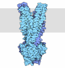
- EMDB-72625: Structure of fimbriae-like lipoprotein by Cryo Electron Microscopy -
+ Open data

- Basic information
| Entry |  | |||||||||
|---|---|---|---|---|---|---|---|---|---|---|
| Title | Structure of fimbriae-like lipoprotein by Cryo Electron Microscopy | |||||||||
 Map data Map data | ||||||||||
 Sample Sample |
| |||||||||
 Keywords Keywords | ffp1 / porphyromonas gingivalis / pili / CELL ADHESION | |||||||||
| Function / homology | Fimbrial subunit protein, C-terminal / Major fimbrial subunit protein type IV, Fimbrillin, C-terminal / Prokaryotic membrane lipoprotein lipid attachment site profile. / Minor fimbrium subunit Mfa1 C-terminal domain-containing protein Function and homology information Function and homology information | |||||||||
| Biological species |  Porphyromonas gingivalis (bacteria) / Porphyromonas gingivalis W83 (bacteria) Porphyromonas gingivalis (bacteria) / Porphyromonas gingivalis W83 (bacteria) | |||||||||
| Method | helical reconstruction / cryo EM / Resolution: 3.2 Å | |||||||||
 Authors Authors | Hanssen E / Gorasia DG / Reynolds EC / Veith PD | |||||||||
| Funding support |  Australia, 1 items Australia, 1 items
| |||||||||
 Citation Citation | Journal: Res Sq / Year: 2026 Title: Novel quadruple helical assembly of a Type V pilin in Porphyromonas gingivalis Authors: Reynolds EC / Gorasia DG / Slakeski N / Gui M / Chen Y-Y / Moore C / Catmull D / Veith P / Dashper S / Hanssen E | |||||||||
| History |
|
- Structure visualization
| Supplemental images |
|---|

- Downloads & links
-EMDB archive
| Map data | emd_72625.map.gz | 15.7 MB | EMDB map data format | |
|---|---|---|---|---|
| Header (meta data) | emd-72625-v30.xmlemd-72625.xml | 18.4 KB 18.4 KB | Display Display | EMDB header |
| FSC (resolution estimation) | emd_72625_fsc.xml | 13.1 KB | Display | FSC data file |
| Images |  emd_72625.png emd_72625.png | 80.2 KB | ||
| Masks | emd_72625_msk_1.map | 244.1 MB | Mask map | |
| Filedesc metadata | emd-72625.cif.gz | 6.5 KB | ||
| Others | emd_72625_half_map_1.map.gzemd_72625_half_map_2.map.gz | 226.4 MB 226.4 MB | ||
| Archive directory |  https://data.pdbj.org/pub/emdb/structures/EMD-72625ftp://data.pdbj.org/pub/emdb/structures/EMD-72625 https://data.pdbj.org/pub/emdb/structures/EMD-72625ftp://data.pdbj.org/pub/emdb/structures/EMD-72625 | HTTPS FTP |
-Related structure data
| Related structure data |  9y6oMC M: atomic model generated by this map C: citing same article ( |
|---|---|
| Similar structure data |
-Links
| EMDB pages | EMDB (EBI/PDBe) / EMDataResource |
|---|---|
| Related items in Molecule of the Month |
-Map
| File | Download / File: emd_72625.map.gz / Format: CCP4 / Size: 244.1 MB / Type: IMAGE STORED AS FLOATING POINT NUMBER (4 BYTES) | ||||||||||||||||||||||||||||||||||||
|---|---|---|---|---|---|---|---|---|---|---|---|---|---|---|---|---|---|---|---|---|---|---|---|---|---|---|---|---|---|---|---|---|---|---|---|---|---|
| Projections & slices | Image control
Images are generated by Spider. | ||||||||||||||||||||||||||||||||||||
| Voxel size | X=Y=Z: 1.32 Å | ||||||||||||||||||||||||||||||||||||
| Density |
| ||||||||||||||||||||||||||||||||||||
| Symmetry | Space group: 1 | ||||||||||||||||||||||||||||||||||||
| Details | EMDB XML:
|
 Z (Sec.)
 Y (Row.)
 X (Col.)


-Supplemental data
-Mask #1
| File | emd_72625_msk_1.map | ||||||||||||
|---|---|---|---|---|---|---|---|---|---|---|---|---|---|
| Projections & Slices |
| ||||||||||||
| Density Histograms |


-Half map: #2
| File | emd_72625_half_map_1.map | ||||||||||||
|---|---|---|---|---|---|---|---|---|---|---|---|---|---|
| Projections & Slices |
| ||||||||||||
| Density Histograms |


-Half map: #1
| File | emd_72625_half_map_2.map | ||||||||||||
|---|---|---|---|---|---|---|---|---|---|---|---|---|---|
| Projections & Slices |
| ||||||||||||
| Density Histograms |


- Sample components
-Entire : ffp1 a type V pilin
| Entire | Name: ffp1 a type V pilin |
|---|---|
| Components |
|
-Supramolecule #1: ffp1 a type V pilin
| Supramolecule | Name: ffp1 a type V pilin / type: complex / ID: 1 / Parent: 0 / Macromolecule list: all |
|---|---|
| Source (natural) | Organism: Porphyromonas gingivalis (bacteria) / Strain: W83 |
-Macromolecule #1: Minor fimbrium subunit Mfa1 C-terminal domain-containing protein
| Macromolecule | Name: Minor fimbrium subunit Mfa1 C-terminal domain-containing protein type: protein_or_peptide / ID: 1 / Number of copies: 2 / Enantiomer: LEVO |
|---|---|
| Source (natural) | Organism: Porphyromonas gingivalis W83 (bacteria) |
| Molecular weight | Theoretical: 46.953746 KDa |
| Sequence | String: KGGTHDPLQS VRRITFLFFH ETDSKLLLSR TVEPTSDLSF DLKIPKQNYR LAVLVNSGSS YAAIIPEILL PTTAIQATSQ TLFESFAAY ETGNITSESE HSVTMANDQG LIKLLSTQIV DKKSQLSEAS RLSVNVEPCL ARVLVVGKPT ISGGEYTGDV S CYVIDVVP ...String: KGGTHDPLQS VRRITFLFFH ETDSKLLLSR TVEPTSDLSF DLKIPKQNYR LAVLVNSGSS YAAIIPEILL PTTAIQATSQ TLFESFAAY ETGNITSESE HSVTMANDQG LIKLLSTQIV DKKSQLSEAS RLSVNVEPCL ARVLVVGKPT ISGGEYTGDV S CYVIDVVP QRIYPLRHLA KLSSGTNEAY GDNSPLADRY ASSWAEESIA AGVAYNNVYG YVKADMFDNP VAATKMQEKK TD FNLNQVA IYTKESTVNP KNYFTAYVPR VVLRAKYVPH GIPGVKPDEG WIEFQGRKMS LEQFKKYVDN PVSAGMALAD SIK KAKADN SLVYTGGFVS HGIQFYYKSQ NYYAIPIRHF DDEKAPNKDS YGRFGLVRNN EYILSVKSIT GAGSPIVPPV STTE AIEKE GYLPASIAVN QTTAHEQDVD L UniProtKB: Minor fimbrium subunit Mfa1 C-terminal domain-containing protein |
-Experimental details
-Structure determination
| Method | cryo EM |
|---|---|
 Processing Processing | helical reconstruction |
| Aggregation state | filament |
-Sample preparation
| Buffer | pH: 7.5 Component:
| |||||||||||||||
|---|---|---|---|---|---|---|---|---|---|---|---|---|---|---|---|---|
| Grid | Model: Quantifoil R1.2/1.3 / Material: COPPER / Pretreatment - Type: GLOW DISCHARGE / Pretreatment - Time: 15 sec. / Pretreatment - Atmosphere: AIR / Details: 15 mA | |||||||||||||||
| Vitrification | Cryogen name: ETHANE / Chamber humidity: 95 % / Chamber temperature: 295 K / Instrument: FEI VITROBOT MARK IV Details: Blot Force -1 Blot time 3 sec temp 22 degres C sample size 4ul. |
- Electron microscopy
| Microscope | TFS KRIOS |
|---|---|
| Image recording | Film or detector model: GATAN K3 BIOQUANTUM (6k x 4k) / Number grids imaged: 1 / Number real images: 8909 / Average exposure time: 5.34 sec. / Average electron dose: 54.0 e/Å2 |
| Electron beam | Acceleration voltage: 300 kV / Electron source:  FIELD EMISSION GUN FIELD EMISSION GUN |
| Electron optics | C2 aperture diameter: 50.0 µm / Illumination mode: FLOOD BEAM / Imaging mode: BRIGHT FIELD / Cs: 2.7 mm / Nominal defocus max: 1.6 µm / Nominal defocus min: 0.6 µm / Nominal magnification: 64000 |
| Sample stage | Specimen holder model: FEI TITAN KRIOS AUTOGRID HOLDER / Cooling holder cryogen: NITROGEN |
| Experimental equipment |  Model: Titan Krios / Image courtesy: FEI Company |


