 Movie
Movie Controller


+ Open data

- Basic information
| Entry | Database: PDB / ID: 9wp9 | ||||||
|---|---|---|---|---|---|---|---|
| Title | Cryo-EM structure of the d18:1 S1P-bound S1PR3 and Gq complex | ||||||
 Components Components |
| ||||||
 Keywords Keywords | MEMBRANE PROTEIN / GPCR / SBDD / lipid | ||||||
| Function / homology |  Function and homology information Function and homology informationnegative regulation of establishment of endothelial barrier / sphingosine-1-phosphate receptor activity / Lysosphingolipid and LPA receptors / regulation of interleukin-1 beta production / anatomical structure morphogenesis / Notch signaling pathway / integrin binding / G protein-coupled receptor activity / adenylate cyclase-inhibiting G protein-coupled receptor signaling pathway / Olfactory Signaling Pathway ...negative regulation of establishment of endothelial barrier / sphingosine-1-phosphate receptor activity / Lysosphingolipid and LPA receptors / regulation of interleukin-1 beta production / anatomical structure morphogenesis / Notch signaling pathway / integrin binding / G protein-coupled receptor activity / adenylate cyclase-inhibiting G protein-coupled receptor signaling pathway / Olfactory Signaling Pathway / Activation of the phototransduction cascade / G protein-coupled acetylcholine receptor signaling pathway / G beta:gamma signalling through PLC beta / Presynaptic function of Kainate receptors / Thromboxane signalling through TP receptor / Activation of G protein gated Potassium channels / Inhibition of voltage gated Ca2+ channels via Gbeta/gamma subunits / G-protein activation / Glucagon signaling in metabolic regulation / G beta:gamma signalling through CDC42 / Prostacyclin signalling through prostacyclin receptor / Synthesis, secretion, and inactivation of Glucagon-like Peptide-1 (GLP-1) / G beta:gamma signalling through BTK / photoreceptor disc membrane / ADP signalling through P2Y purinoceptor 12 / Glucagon-type ligand receptors / Sensory perception of sweet, bitter, and umami (glutamate) taste / Adrenaline,noradrenaline inhibits insulin secretion / Vasopressin regulates renal water homeostasis via Aquaporins / Glucagon-like Peptide-1 (GLP1) regulates insulin secretion / G alpha (z) signalling events / cellular response to catecholamine stimulus / ADP signalling through P2Y purinoceptor 1 / G beta:gamma signalling through PI3Kgamma / ADORA2B mediated anti-inflammatory cytokines production / adenylate cyclase-activating dopamine receptor signaling pathway / Cooperation of PDCL (PhLP1) and TRiC/CCT in G-protein beta folding / GPER1 signaling / cellular response to prostaglandin E stimulus / heterotrimeric G-protein complex / G alpha (12/13) signalling events / Inactivation, recovery and regulation of the phototransduction cascade / G-protein beta-subunit binding / extracellular vesicle / sensory perception of taste / presynapse / Thrombin signalling through proteinase activated receptors (PARs) / adenylate cyclase-activating G protein-coupled receptor signaling pathway / signaling receptor complex adaptor activity / positive regulation of cytosolic calcium ion concentration / retina development in camera-type eye / fibroblast proliferation / GTPase binding / Ca2+ pathway / High laminar flow shear stress activates signaling by PIEZO1 and PECAM1:CDH5:KDR in endothelial cells / G alpha (i) signalling events / G alpha (s) signalling events / G alpha (q) signalling events / phospholipase C-activating G protein-coupled receptor signaling pathway / Ras protein signal transduction / Extra-nuclear estrogen signaling / cell population proliferation / G protein-coupled receptor signaling pathway / inflammatory response / lysosomal membrane / GTPase activity / positive regulation of cell population proliferation / lipid binding / synapse / protein-containing complex binding / signal transduction / extracellular exosome / membrane / plasma membrane / cytoplasm / cytosol Similarity search - Function | ||||||
| Biological species |  Homo sapiens (human) Homo sapiens (human) | ||||||
| Method | ELECTRON MICROSCOPY / single particle reconstruction / cryo EM / Resolution: 3.25 Å | ||||||
 Authors Authors | Im, D. / Asada, H. / Iwata, S. / Yamauchi, M. / Hagiwara, M. | ||||||
| Funding support | 1items
| ||||||
 Citation Citation | Journal: Proc Natl Acad Sci U S A / Year: 2025 Title: Structural insights into the G-protein subtype selectivity revealed by human sphingosine-1-phosphate receptor 3-G complexes. Authors: Momono Yamauchi / Dohyun Im / Shintaro Maeda / Tatsuya Ikuta / Masayasu Toyomoto / Hidetsugu Asada / Yukihiko Sugita / Jun-Ichi Kishikawa / Takeshi Noda / Takayuki Kato / Asuka Inoue / So ...Authors: Momono Yamauchi / Dohyun Im / Shintaro Maeda / Tatsuya Ikuta / Masayasu Toyomoto / Hidetsugu Asada / Yukihiko Sugita / Jun-Ichi Kishikawa / Takeshi Noda / Takayuki Kato / Asuka Inoue / So Iwata / Masatoshi Hagiwara /  Abstract: Sphingosine-1-phosphate (S1P) is one of the most extensively studied bioactive lipids that transduces signals via the S1P receptor (S1PR) family (S1PR1-5), a class of G-protein-coupled receptors ...Sphingosine-1-phosphate (S1P) is one of the most extensively studied bioactive lipids that transduces signals via the S1P receptor (S1PR) family (S1PR1-5), a class of G-protein-coupled receptors (GPCRs), to regulate immune cell migration, vascular permeability, and pain modulation. However, the mechanism for achieving specificity in downstream signaling remains poorly understood. Here, we present cryogenic electron microscopic structures of the S1PR3-G complex bound to endogenous agonists: d18:1 S1P or d16:1 S1P. Both agonists shared the same binding pocket and binding mode despite the different signaling intensities of the S1PR3-G signal pathway. By comparing the structures of two agonist-bound complexes, combined with mutagenesis studies, we identified key amino acids, Phe119 and Arg136, that play crucial roles in differential agonist recognition and receptor activation. Furthermore, structural comparisons with previously determined S1PR3-G complex or G-protein-free S1PR3 structures, along with mutagenesis analysis, revealed dynamic intracellular loop 2 conformations and specific amino acid interactions that contribute to G-protein selectivity. Notably, we identified amino acids at the 34.50 and 34.53 positions within ICL2 as critical for specific interactions with G proteins. These findings provide better understanding of the mechanism of GPCR activation and unique perspectives that can be applied to other class A GPCRs, leading to the possibility of optimized drug development. | ||||||
| History |
|
- Structure visualization
| Structure viewer | Molecule: MolmilJmol/JSmol |
|---|
- Downloads & links
-Download
| PDBx/mmCIF format | 9wp9.cif.gz | 259 KB | Display | PDBx/mmCIF format |
|---|---|---|---|---|
| PDB format | pdb9wp9.ent.gz | 201.1 KB | Display | PDB format |
| PDBx/mmJSON format | 9wp9.json.gz | Tree view | PDBx/mmJSON format | |
| Others |  Other downloads Other downloads |
-Validation report
| Arichive directory | https://data.pdbj.org/pub/pdb/validation_reports/wp/9wp9ftp://data.pdbj.org/pub/pdb/validation_reports/wp/9wp9 | HTTPS FTP |
|---|
-Related structure data
| Related structure data |  66136MC  9l74C M: map data used to model this data C: citing same article ( |
|---|---|
| Similar structure data |

-Links
 PDBj
PDBj


- Assembly
| Deposited unit | 
|
|---|---|
| 1 |
|
-Components
-Protein , 3 types, 3 molecules ANR
| #1: Protein | Mass: 27680.387 Da / Num. of mol.: 1 Source method: isolated from a genetically manipulated source Source: (gene. exp.) Homo sapiens (human) / Production host:   Spodoptera frugiperda (fall armyworm) Spodoptera frugiperda (fall armyworm) |
|---|---|
| #5: Protein | Mass: 14016.636 Da / Num. of mol.: 1 Source method: isolated from a genetically manipulated source Source: (gene. exp.) Homo sapiens (human) / Production host: Spodoptera frugiperda (fall armyworm) |
| #6: Protein | Mass: 38829.141 Da / Num. of mol.: 1 Source method: isolated from a genetically manipulated source Source: (gene. exp.) Homo sapiens (human) / Gene: S1PR3, C9orf108, C9orf47, EDG3 / Production host: Spodoptera frugiperda (fall armyworm) / References: UniProt: Q99500 |
-Guanine nucleotide-binding protein ... , 2 types, 2 molecules BG
| #2: Protein | Mass: 39418.086 Da / Num. of mol.: 1 Source method: isolated from a genetically manipulated source Source: (gene. exp.) Homo sapiens (human) / Gene: GNB1 / Production host: Spodoptera frugiperda (fall armyworm) / References: UniProt: P62873 |
|---|---|
| #4: Protein | Mass: 7861.143 Da / Num. of mol.: 1 Source method: isolated from a genetically manipulated source Source: (gene. exp.) Homo sapiens (human) / Gene: GNG2 / Production host: Spodoptera frugiperda (fall armyworm) / References: UniProt: P59768 |
-Antibody / Non-polymers , 2 types, 2 molecules C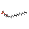
| #3: Antibody | Mass: 27784.896 Da / Num. of mol.: 1 Source method: isolated from a genetically manipulated source Source: (gene. exp.) Homo sapiens (human) / Production host: Spodoptera frugiperda (fall armyworm) |
|---|---|
| #7: Chemical | ChemComp-S1P / ( Mass: 379.472 Da / Num. of mol.: 1 / Source method: obtained synthetically / Formula: C18H38NO5P / Feature type: SUBJECT OF INVESTIGATION Mass: 379.472 Da / Num. of mol.: 1 / Source method: obtained synthetically / Formula: C18H38NO5P / Feature type: SUBJECT OF INVESTIGATION |
-Details
| Has ligand of interest | Y |
|---|---|
| Has protein modification | Y |
-Experimental details
-Experiment
| Experiment | Method: ELECTRON MICROSCOPY |
|---|---|
| EM experiment | Aggregation state: PARTICLE / 3D reconstruction method: single particle reconstruction |
- Sample preparation
| Component | Name: d18:1 S1P-bound S1PR3 in complex with Gq / Type: COMPLEX / Entity ID: #1-#6 / Source: RECOMBINANT |
|---|---|
| Molecular weight | Value: 0.19 MDa / Experimental value: YES |
| Source (natural) | Organism: Homo sapiens (human) |
| Source (recombinant) | Organism: Spodoptera frugiperda (fall armyworm) |
| Buffer solution | pH: 7.5 |
| Specimen | Conc.: 10 mg/ml / Embedding applied: NO / Shadowing applied: NO / Staining applied: NO / Vitrification applied: YES |
| Vitrification | Cryogen name: ETHANE / Humidity: 100 % / Chamber temperature: 281.15 K |
- Electron microscopy imaging
| Experimental equipment |  Model: Titan Krios / Image courtesy: FEI Company |
|---|---|
| Microscopy | Model: TFS KRIOS |
| Electron gun | Electron source:  FIELD EMISSION GUN / Accelerating voltage: 300 kV / Illumination mode: FLOOD BEAM FIELD EMISSION GUN / Accelerating voltage: 300 kV / Illumination mode: FLOOD BEAM |
| Electron lens | Mode: BRIGHT FIELD / Nominal defocus max: 1800 nm / Nominal defocus min: 800 nm |
| Specimen holder | Specimen holder model: FEI TITAN KRIOS AUTOGRID HOLDER |
| Image recording | Electron dose: 50 e/Å2 / Film or detector model: GATAN K3 BIOQUANTUM (6k x 4k) |
- Processing
| EM software |
| ||||||||||||||||||||||||
|---|---|---|---|---|---|---|---|---|---|---|---|---|---|---|---|---|---|---|---|---|---|---|---|---|---|
| CTF correction | Type: NONE | ||||||||||||||||||||||||
| 3D reconstruction | Resolution: 3.25 Å / Resolution method: FSC 0.143 CUT-OFF / Num. of particles: 366554 / Symmetry type: POINT | ||||||||||||||||||||||||
| Refinement | Highest resolution: 3.25 Å Stereochemistry target values: REAL-SPACE (WEIGHTED MAP SUM AT ATOM CENTERS) | ||||||||||||||||||||||||
| Refine LS restraints |
|
