 Movie
Movie Controller

[English] 日本語
 Yorodumi
- PDB-9wno: Cryo-EM structure of Candida glabrata GPI mannosyltransferase I b... -
+ Open data

- Basic information
| Entry | Database: PDB / ID: 9wno | ||||||
|---|---|---|---|---|---|---|---|
| Title | Cryo-EM structure of Candida glabrata GPI mannosyltransferase I bound to Dol-P-Man | ||||||
 Components Components |
| ||||||
 Keywords Keywords | MEMBRANE PROTEIN / GT-C / GPI / Mannosyltransferase | ||||||
| Function / homology |  Function and homology information Function and homology informationdol-P-Man:GlcN-acyl-PI alpha-1,4-mannosyltransferase activity / glycosylphosphatidylinositol-mannosyltransferase I complex / mannosyltransferase activity / GPI anchor biosynthetic process / fungal-type cell wall organization / Transferases; Glycosyltransferases; Hexosyltransferases / ERAD pathway / protein processing / endoplasmic reticulum membrane Similarity search - Function | ||||||
| Biological species |  Nakaseomyces glabratus (fungus) Nakaseomyces glabratus (fungus) | ||||||
| Method | ELECTRON MICROSCOPY / single particle reconstruction / cryo EM / Resolution: 3.48 Å | ||||||
 Authors Authors | Sun, H. / Wu, W.H. / Yan, Z.F. | ||||||
| Funding support | 1items
| ||||||
 Citation Citation | Journal: J Fungi (Basel) / Year: 2025 Title: Structural Insights into the Glycosylphosphatidylinositol Mannosyltransferase I Complex from . Authors: Hui Sun / Weihong Wu / Xiaomei Li / Yang Deng / Jiarong Huang / Meng Yin / Zhaofeng Yan /  Abstract: The global rise in resistance to first-line antifungal agents highlights the urgent need for new therapeutic strategies. Glycosylphosphatidylinositol (GPI)-anchored protein biosynthesis is an ...The global rise in resistance to first-line antifungal agents highlights the urgent need for new therapeutic strategies. Glycosylphosphatidylinositol (GPI)-anchored protein biosynthesis is an attractive target. The GPI mannosyltransferase I (GPI-MT-I), composed of Gpi14 and Pbn1, catalyzes the essential first mannose transfer from dolichol-phosphomannose (Dol-P-Man) to the GPI precursor. This initial mannosylation is critical for fungal cell wall integrity, yet the molecular basis of GPI-MT-I assembly and substrate recognition remains poorly understood. Here, we present the cryo-EM structure of GPI-MT-I in complex with Dol-P-Man, revealing how Gpi14 and Pbn1 form a stable complex and engage the mannose donor. An AlphaFold3-predicted acceptor-bound model further defines the structural basis of acceptor substrate recognition and suggests a plausible catalytic mechanism. Comparison with structural homologs highlights a distinct mode of substrate engagement by GPI-MT-I. Together, these findings establish a mechanistic framework for GPI-MT-I function with broader implications for the GPI-MT family. | ||||||
| History |
|
- Structure visualization
| Structure viewer | Molecule: MolmilJmol/JSmol |
|---|
- Downloads & links
-Download
| PDBx/mmCIF format | 9wno.cif.gz | 185.5 KB | Display | PDBx/mmCIF format |
|---|---|---|---|---|
| PDB format | pdb9wno.ent.gz | 141 KB | Display | PDB format |
| PDBx/mmJSON format | 9wno.json.gz | Tree view | PDBx/mmJSON format | |
| Others |  Other downloads Other downloads |
-Validation report
| Arichive directory | https://data.pdbj.org/pub/pdb/validation_reports/wn/9wnoftp://data.pdbj.org/pub/pdb/validation_reports/wn/9wno | HTTPS FTP |
|---|
-Related structure data
| Related structure data |  66120MC M: map data used to model this data C: citing same article ( |
|---|---|
| Similar structure data |
-Links
 PDBj
PDBj- Assembly
| Deposited unit | 
|
|---|---|
| 1 |
|
-Components
-Protein , 2 types, 2 molecules AB
| #1: Protein | Mass: 50289.254 Da / Num. of mol.: 1 Source method: isolated from a genetically manipulated source Details: Sequence reference for strain 'Nakaseomyces glabratus' is not available in UniProt at the time of biocuration. Current sequence reference is from UniProt id Q6FXQ5. Source: (gene. exp.) Nakaseomyces glabratus (fungus) / Gene: GPI14, CAGL0B03905g / Production host:  References: UniProt: Q6FXQ5, Transferases; Glycosyltransferases; Hexosyltransferases |
|---|---|
| #2: Protein | Mass: 47799.812 Da / Num. of mol.: 1 Source method: isolated from a genetically manipulated source Details: Sequence reference for strain 'Nakaseomyces glabratus' is not available in UniProt at the time of biocuration. Current sequence reference is from UniProt id Q6FX62. Source: (gene. exp.) Nakaseomyces glabratus (fungus) / Gene: PBN1, CAGL0B00506g / Production host: |
-Sugars , 2 types, 3 molecules 
| #3: Polysaccharide | Source method: isolated from a genetically manipulated source #6: Sugar | ChemComp-NAG / |  Type: D-saccharide, beta linking / Mass: 221.208 Da / Num. of mol.: 1 / Source method: obtained synthetically / Formula: C8H15NO6 / Feature type: SUBJECT OF INVESTIGATION Type: D-saccharide, beta linking / Mass: 221.208 Da / Num. of mol.: 1 / Source method: obtained synthetically / Formula: C8H15NO6 / Feature type: SUBJECT OF INVESTIGATION |
|---|
-Non-polymers , 2 types, 2 molecules 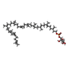

| #4: Chemical | ChemComp-MJC /  Mass: 1011.439 Da / Num. of mol.: 1 / Source method: obtained synthetically / Formula: C61H103O9P / Feature type: SUBJECT OF INVESTIGATION Mass: 1011.439 Da / Num. of mol.: 1 / Source method: obtained synthetically / Formula: C61H103O9P / Feature type: SUBJECT OF INVESTIGATION |
|---|---|
| #5: Chemical | ChemComp-Y01 /  Mass: 486.726 Da / Num. of mol.: 1 / Source method: obtained synthetically / Formula: C31H50O4 / Feature type: SUBJECT OF INVESTIGATION Mass: 486.726 Da / Num. of mol.: 1 / Source method: obtained synthetically / Formula: C31H50O4 / Feature type: SUBJECT OF INVESTIGATION |
-Details
| Has ligand of interest | Y |
|---|---|
| Has protein modification | Y |
-Experimental details
-Experiment
| Experiment | Method: ELECTRON MICROSCOPY |
|---|---|
| EM experiment | Aggregation state: PARTICLE / 3D reconstruction method: single particle reconstruction |
- Sample preparation
| Component | Name: GPI mannosyltransferase I / Type: COMPLEX / Entity ID: #1-#2 / Source: RECOMBINANT |
|---|---|
| Source (natural) | Organism: Nakaseomyces glabratus (fungus) |
| Source (recombinant) | Organism: |
| Buffer solution | pH: 7.4 |
| Specimen | Embedding applied: NO / Shadowing applied: NO / Staining applied: NO / Vitrification applied: YES |
| Vitrification | Cryogen name: ETHANE |
- Electron microscopy imaging
| Experimental equipment |  Model: Titan Krios / Image courtesy: FEI Company |
|---|---|
| Microscopy | Model: TFS KRIOS |
| Electron gun | Electron source:  FIELD EMISSION GUN / Accelerating voltage: 300 kV / Illumination mode: FLOOD BEAM FIELD EMISSION GUN / Accelerating voltage: 300 kV / Illumination mode: FLOOD BEAM |
| Electron lens | Mode: BRIGHT FIELD / Nominal defocus max: 2100 nm / Nominal defocus min: 1700 nm |
| Image recording | Electron dose: 56 e/Å2 / Film or detector model: GATAN K3 (6k x 4k) |
- Processing
| EM software |
| ||||||||||||||||||||||||
|---|---|---|---|---|---|---|---|---|---|---|---|---|---|---|---|---|---|---|---|---|---|---|---|---|---|
| CTF correction | Type: PHASE FLIPPING AND AMPLITUDE CORRECTION | ||||||||||||||||||||||||
| 3D reconstruction | Resolution: 3.48 Å / Resolution method: FSC 0.143 CUT-OFF / Num. of particles: 80856 / Symmetry type: POINT | ||||||||||||||||||||||||
| Refinement | Highest resolution: 3.48 Å Stereochemistry target values: REAL-SPACE (WEIGHTED MAP SUM AT ATOM CENTERS) | ||||||||||||||||||||||||
| Refine LS restraints |
|
