 Movie
Movie Controller


+ Open data

- Basic information
| Entry | Database: PDB / ID: 9w54 | ||||||
|---|---|---|---|---|---|---|---|
| Title | Structure of L-glutamate oxidase in complex with L-glutamate | ||||||
 Components Components | (L-glutamate oxidase ...) x 3 | ||||||
 Keywords Keywords | OXIDOREDUCTASE / L-glutamic acid oxidase | ||||||
| Function / homology |  Function and homology information Function and homology informationL-glutamate oxidase / L-amino-acid oxidase activity / amino acid catabolic process / nucleotide binding / extracellular region Similarity search - Function | ||||||
| Biological species |  Streptomyces sp. X-119-6 (bacteria) Streptomyces sp. X-119-6 (bacteria) | ||||||
| Method |  X-RAY DIFFRACTION / SYNCHROTRON / MOLECULAR REPLACEMENT / Resolution: 2.55 Å X-RAY DIFFRACTION / SYNCHROTRON / MOLECULAR REPLACEMENT / Resolution: 2.55 Å | ||||||
 Authors Authors | Ueda, Y. / Takekawa, N. / Nakayama, N. / Inagaki, K. / Imada, K. | ||||||
| Funding support |  Japan, 1items Japan, 1items
| ||||||
 Citation Citation | Journal: Protein Sci. / Year: 2026 Title: Substrate recognition mechanisms of ʟ-glutamate oxidase from Streptomyces sp. and its conversion to ʟ-tyrosine oxidase. Authors: Ueda, Y. / Yano, Y. / Nakayama, N. / Takekawa, N. / Inagaki, K. / Imada, K. | ||||||
| History |
|
- Structure visualization
| Structure viewer | Molecule: MolmilJmol/JSmol |
|---|
- Downloads & links
-Download
| PDBx/mmCIF format | 9w54.cif.gz | 155.2 KB | Display | PDBx/mmCIF format |
|---|---|---|---|---|
| PDB format | pdb9w54.ent.gz | 108.2 KB | Display | PDB format |
| PDBx/mmJSON format | 9w54.json.gz | Tree view | PDBx/mmJSON format | |
| Others |  Other downloads Other downloads |
-Validation report
| Arichive directory | https://data.pdbj.org/pub/pdb/validation_reports/w5/9w54ftp://data.pdbj.org/pub/pdb/validation_reports/w5/9w54 | HTTPS FTP |
|---|
-Related structure data
| Related structure data |  9w55C  9w56C  9w57C  9w58C 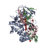 2e1mS S: Starting model for refinement C: citing same article ( |
|---|---|
| Similar structure data |
-Links
 PDBj
PDBj


- Assembly
| Deposited unit | 
| ||||||||||
|---|---|---|---|---|---|---|---|---|---|---|---|
| 1 | 
| ||||||||||
| Unit cell |
| ||||||||||
| Components on special symmetry positions |
|
-Components
-L-glutamate oxidase ... , 3 types, 3 molecules ABC
| #1: Protein | Mass: 42019.227 Da / Num. of mol.: 1 Source method: isolated from a genetically manipulated source Details: The L-glutamate oxidase alpha fragment (A15-Y390) produced by proteolytic processing of the L-glutamate oxidase precursor (uniprot ID:Q8L3C7). Source: (gene. exp.) Streptomyces sp. X-119-6 (bacteria) / Strain: X-119-6 / Gene: lgoX / Plasmid: pMal-c2 / Production host: |
|---|---|
| #2: Protein | Mass: 10575.776 Da / Num. of mol.: 1 Source method: isolated from a genetically manipulated source Details: The L-glutamate oxidase gamma fragment (A391-A480) produced by proteolytic processing of the L-glutamate oxidase precursor (uniprot ID:Q8L3C7). Source: (gene. exp.) Streptomyces sp. X-119-6 (bacteria) / Strain: X-119-6 / Gene: lgoX / Plasmid: pMal-c2 / Production host: |
| #3: Protein | Mass: 17452.059 Da / Num. of mol.: 1 Source method: isolated from a genetically manipulated source Details: The L-glutamate oxidase beta fragment (G521-G683) produced by proteolytic processing of the L-glutamate oxidase precursor (uniprot ID:Q8L3C7). Source: (gene. exp.) Streptomyces sp. X-119-6 (bacteria) / Strain: X-119-6 / Gene: lgoX / Plasmid: pMal-c2 / Production host: |
-Non-polymers , 3 types, 221 molecules 


| #4: Chemical | ChemComp-FAD /  Mass: 785.550 Da / Num. of mol.: 1 / Source method: obtained synthetically / Formula: C27H33N9O15P2 / Comment: FAD*YM Mass: 785.550 Da / Num. of mol.: 1 / Source method: obtained synthetically / Formula: C27H33N9O15P2 / Comment: FAD*YM |
|---|---|
| #5: Chemical | ChemComp-GLU /  Type: L-peptide linking / Mass: 147.129 Da / Num. of mol.: 1 / Source method: obtained synthetically / Formula: C5H9NO4 / Feature type: SUBJECT OF INVESTIGATION Type: L-peptide linking / Mass: 147.129 Da / Num. of mol.: 1 / Source method: obtained synthetically / Formula: C5H9NO4 / Feature type: SUBJECT OF INVESTIGATION |
| #6: Water | ChemComp-HOH / Mass: 18.015 Da / Num. of mol.: 219 / Source method: isolated from a natural source / Formula: H2O |
-Details
| Has ligand of interest | Y |
|---|---|
| Has protein modification | N |
-Experimental details
-Experiment
| Experiment | Method: X-RAY DIFFRACTION / Number of used crystals: 1 |
|---|
- Sample preparation
| Crystal | Density Matthews: 2.69 Å3/Da / Density % sol: 54.23 % |
|---|---|
| Crystal grow | Temperature: 293 K / Method: vapor diffusion, sitting drop Details: 0.1 M CAPS pH10, 1.2 M NaH2PO4, 0.8 M K2HPO4, 0.2 M Li2SO4 |
-Data collection
| Diffraction | Mean temperature: 100 K / Serial crystal experiment: N |
|---|---|
| Diffraction source | Source: SYNCHROTRON / Site: SPring-8 / Beamline: BL41XU / Wavelength: 1 Å |
| Detector | Type: DECTRIS EIGER X 16M / Detector: PIXEL / Date: Apr 19, 2022 |
| Radiation | Monochromator: Double-crystal monochromator / Protocol: SINGLE WAVELENGTH / Monochromatic (M) / Laue (L): M / Scattering type: x-ray |
| Radiation wavelength | Wavelength: 1 Å / Relative weight: 1 |
| Reflection | Resolution: 2.55→62.16 Å / Num. obs: 25482 / % possible obs: 99.2 % / Redundancy: 3.8 % / CC1/2: 0.967 / Net I/σ(I): 8.2 |
| Reflection shell | Resolution: 2.55→2.66 Å / Redundancy: 3.9 % / Mean I/σ(I) obs: 2.2 / Num. unique obs: 3022 / CC1/2: 0.72 / % possible all: 98.9 |
- Processing
| Software |
| |||||||||||||||||||||||||||||||||||||||||||||||||||||||||||||||||||||||||||||||||||||||||||||||||||||||||
|---|---|---|---|---|---|---|---|---|---|---|---|---|---|---|---|---|---|---|---|---|---|---|---|---|---|---|---|---|---|---|---|---|---|---|---|---|---|---|---|---|---|---|---|---|---|---|---|---|---|---|---|---|---|---|---|---|---|---|---|---|---|---|---|---|---|---|---|---|---|---|---|---|---|---|---|---|---|---|---|---|---|---|---|---|---|---|---|---|---|---|---|---|---|---|---|---|---|---|---|---|---|---|---|---|---|---|
| Refinement | Method to determine structure: MOLECULAR REPLACEMENT Starting model: 2E1M Resolution: 2.55→62.15 Å / SU ML: 0.3324 / Cross valid method: FREE R-VALUE / σ(F): 1.35 / Phase error: 23.8092 Stereochemistry target values: GeoStd + Monomer Library + CDL v1.2
| |||||||||||||||||||||||||||||||||||||||||||||||||||||||||||||||||||||||||||||||||||||||||||||||||||||||||
| Solvent computation | Shrinkage radii: 0.9 Å / VDW probe radii: 1.1 Å / Solvent model: FLAT BULK SOLVENT MODEL | |||||||||||||||||||||||||||||||||||||||||||||||||||||||||||||||||||||||||||||||||||||||||||||||||||||||||
| Displacement parameters | Biso mean: 37.41 Å2 | |||||||||||||||||||||||||||||||||||||||||||||||||||||||||||||||||||||||||||||||||||||||||||||||||||||||||
| Refinement step | Cycle: LAST / Resolution: 2.55→62.15 Å
| |||||||||||||||||||||||||||||||||||||||||||||||||||||||||||||||||||||||||||||||||||||||||||||||||||||||||
| Refine LS restraints |
| |||||||||||||||||||||||||||||||||||||||||||||||||||||||||||||||||||||||||||||||||||||||||||||||||||||||||
| LS refinement shell |
|
