[English] 日本語
 Yorodumi
- PDB-9v7k: Phycobilisome allophycocyanin hexamer C from Gloeobacter violaceu... -
+ Open data
- Basic information
| Entry | Database: PDB / ID: 9v7k | |||||||||||||||||||||
|---|---|---|---|---|---|---|---|---|---|---|---|---|---|---|---|---|---|---|---|---|---|---|
| Title | Phycobilisome allophycocyanin hexamer C from Gloeobacter violaceus PCC 7421 | |||||||||||||||||||||
 Components Components |
| |||||||||||||||||||||
 Keywords Keywords | PHOTOSYNTHESIS / Phycobilisome | |||||||||||||||||||||
| Function / homology |  Function and homology information Function and homology informationphycobilisome / plasma membrane-derived thylakoid membrane / photosynthesis / lyase activity Similarity search - Function | |||||||||||||||||||||
| Biological species |  Gloeobacter violaceus PCC 7421 (bacteria) Gloeobacter violaceus PCC 7421 (bacteria) | |||||||||||||||||||||
| Method | ELECTRON MICROSCOPY / single particle reconstruction / cryo EM / Resolution: 2.94 Å | |||||||||||||||||||||
 Authors Authors | Burtseva, A.D. / Baymukhametov, T.N. / Slonimskiy, Y.B. / Popov, V.O. / Sluchanko, N.N. / Boyko, K.M. | |||||||||||||||||||||
| Funding support |  Russian Federation, 1items Russian Federation, 1items
| |||||||||||||||||||||
 Citation Citation |  Journal: Sci Adv / Year: 2025 Journal: Sci Adv / Year: 2025Title: Structure and quenching of a bundle-shaped phycobilisome. Authors: Anna D Burtseva / Yury B Slonimskiy / Timur N Baymukhametov / Maria A Sinetova / Daniil A Gvozdev / Georgy V Tsoraev / Dmitry A Cherepanov / Eugene G Maksimov / Vladimir O Popov / Konstantin ...Authors: Anna D Burtseva / Yury B Slonimskiy / Timur N Baymukhametov / Maria A Sinetova / Daniil A Gvozdev / Georgy V Tsoraev / Dmitry A Cherepanov / Eugene G Maksimov / Vladimir O Popov / Konstantin M Boyko / Nikolai N Sluchanko /  Abstract: Cyanobacteria use soluble antenna megacomplexes, phycobilisomes (PBSs), to maximize light-harvesting efficiency and small photoswitchable orange carotenoid proteins (OCPs) to down-regulate PBSs in ...Cyanobacteria use soluble antenna megacomplexes, phycobilisomes (PBSs), to maximize light-harvesting efficiency and small photoswitchable orange carotenoid proteins (OCPs) to down-regulate PBSs in high light. Among known PBS morphologies, the one from the basal cyanobacterial genus still lacks detailed structural characterization. Here, we reconstructed a cryo-electron microscopy structure of the >10-megadalton PBS, with diverging, conformationally mobile bundles of rods composed of stacked phycoerythrin and phycocyanin hexamers, stemming from a pentacylindrical allophycocyanin core belted by auxiliary phycocyanin hexamers. We show how two -specific multidomain linker proteins, Glr1262 and Glr2806, maintain this bundle-shaped architecture and reveal its differential regulation via nonphotochemical quenching by two OCP types of that recognize separate binding sites within the allophycocyanin core, including lateral cylinders absent in tricylindrical cores. | |||||||||||||||||||||
| History |
|
- Structure visualization
| Structure viewer | Molecule:  Molmil Molmil Jmol/JSmol Jmol/JSmol |
|---|
- Downloads & links
- Download
| PDBx/mmCIF format |  9v7k.cif.gz 9v7k.cif.gz | 485.7 KB | Display |  PDBx/mmCIF format PDBx/mmCIF format |
|---|---|---|---|---|
| PDB format |  pdb9v7k.ent.gz pdb9v7k.ent.gz | 371.7 KB | Display |  PDB format PDB format |
| PDBx/mmJSON format |  9v7k.json.gz 9v7k.json.gz | Tree view |  PDBx/mmJSON format PDBx/mmJSON format | |
| Others |  Other downloads Other downloads |
-Validation report
| Arichive directory |  https://data.pdbj.org/pub/pdb/validation_reports/v7/9v7k https://data.pdbj.org/pub/pdb/validation_reports/v7/9v7k ftp://data.pdbj.org/pub/pdb/validation_reports/v7/9v7k ftp://data.pdbj.org/pub/pdb/validation_reports/v7/9v7k | HTTPS FTP |
|---|
-Related structure data
| Related structure data |  64816MC  9v7gC  9v7hC  9v7iC  9v7jC  9v7lC M: map data used to model this data C: citing same article ( |
|---|---|
| Similar structure data | Similarity search - Function & homology  F&H Search F&H Search |
- Links
Links
- Assembly
| Deposited unit | 
|
|---|---|
| 1 |
|
- Components
-Protein , 4 types, 4 molecules 1ZMX
| #1: Protein | Mass: 130002.258 Da / Num. of mol.: 1 / Source method: isolated from a natural source / Source: (natural)  Gloeobacter violaceus PCC 7421 (bacteria) / References: UniProt: Q7NL81 Gloeobacter violaceus PCC 7421 (bacteria) / References: UniProt: Q7NL81 |
|---|---|
| #2: Protein | Mass: 92127.602 Da / Num. of mol.: 1 / Source method: isolated from a natural source / Source: (natural)  Gloeobacter violaceus PCC 7421 (bacteria) / References: UniProt: Q7NL64 Gloeobacter violaceus PCC 7421 (bacteria) / References: UniProt: Q7NL64 |
| #5: Protein | Mass: 7765.004 Da / Num. of mol.: 1 / Source method: isolated from a natural source / Source: (natural)  Gloeobacter violaceus PCC 7421 (bacteria) / References: UniProt: Q7NL78 Gloeobacter violaceus PCC 7421 (bacteria) / References: UniProt: Q7NL78 |
| #6: Protein | Mass: 81544.312 Da / Num. of mol.: 1 / Source method: isolated from a natural source / Source: (natural)  Gloeobacter violaceus PCC 7421 (bacteria) / References: UniProt: Q7NGT2 Gloeobacter violaceus PCC 7421 (bacteria) / References: UniProt: Q7NGT2 |
-Allophycocyanin ... , 2 types, 12 molecules ACEGIKBDFHJL
| #3: Protein | Mass: 17555.062 Da / Num. of mol.: 6 / Source method: isolated from a natural source / Source: (natural)  Gloeobacter violaceus PCC 7421 (bacteria) / References: UniProt: Q7NL80 Gloeobacter violaceus PCC 7421 (bacteria) / References: UniProt: Q7NL80#4: Protein | Mass: 17241.664 Da / Num. of mol.: 6 / Source method: isolated from a natural source / Source: (natural)  Gloeobacter violaceus PCC 7421 (bacteria) / References: UniProt: Q7NL79 Gloeobacter violaceus PCC 7421 (bacteria) / References: UniProt: Q7NL79 |
|---|
-Non-polymers , 1 types, 12 molecules 
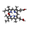
| #7: Chemical | ChemComp-CYC / |
|---|
-Details
| Has ligand of interest | Y |
|---|---|
| Has protein modification | Y |
-Experimental details
-Experiment
| Experiment | Method: ELECTRON MICROSCOPY |
|---|---|
| EM experiment | Aggregation state: PARTICLE / 3D reconstruction method: single particle reconstruction |
- Sample preparation
| Component | Name: Bundle-shaped phycobilisome / Type: COMPLEX / Entity ID: #1-#6 / Source: NATURAL |
|---|---|
| Source (natural) | Organism:  Gloeobacter violaceus PCC 7421 (bacteria) Gloeobacter violaceus PCC 7421 (bacteria) |
| Buffer solution | pH: 7 |
| Buffer component | Conc.: 50 mM / Name: Tris(hydroxymethyl)aminomethane hydrochloride / Formula: Tris-HCl |
| Specimen | Embedding applied: NO / Shadowing applied: NO / Staining applied: NO / Vitrification applied: YES |
| Vitrification | Cryogen name: ETHANE |
- Electron microscopy imaging
| Experimental equipment |  Model: Titan Krios / Image courtesy: FEI Company |
|---|---|
| Microscopy | Model: TFS KRIOS / Details: Preliminary grid screening was performed manually. |
| Electron gun | Electron source:  FIELD EMISSION GUN / Accelerating voltage: 300 kV / Illumination mode: FLOOD BEAM FIELD EMISSION GUN / Accelerating voltage: 300 kV / Illumination mode: FLOOD BEAM |
| Electron lens | Mode: BRIGHT FIELD / Nominal magnification: 85000 X / Nominal defocus max: 1600 nm / Nominal defocus min: 600 nm / Cs: 0.01 mm / C2 aperture diameter: 100 µm / Alignment procedure: ZEMLIN TABLEAU |
| Specimen holder | Cryogen: NITROGEN / Specimen holder model: FEI TITAN KRIOS AUTOGRID HOLDER |
| Image recording | Average exposure time: 3.9 sec. / Electron dose: 66 e/Å2 / Film or detector model: GATAN K3 BIOQUANTUM (6k x 4k) |
| EM imaging optics | Energyfilter name: GIF Bioquantum / Energyfilter slit width: 20 eV Spherical aberration corrector: Microscope was modified with a Cs corrector (CEOS GmbH, Germany). |
| Image scans | Width: 5760 / Height: 4092 |
- Processing
| EM software |
| ||||||||||||||||||||
|---|---|---|---|---|---|---|---|---|---|---|---|---|---|---|---|---|---|---|---|---|---|
| CTF correction | Type: NONE | ||||||||||||||||||||
| Symmetry | Point symmetry: C1 (asymmetric) | ||||||||||||||||||||
| 3D reconstruction | Resolution: 2.94 Å / Resolution method: FSC 0.143 CUT-OFF / Num. of particles: 746972 / Symmetry type: POINT |
 Movie
Movie Controller


 PDBj
PDBj
