 Movie
Movie Controller


+ Open data

- Basic information
| Entry | Database: PDB / ID: 9v75 | |||||||||||||||||||||||||||
|---|---|---|---|---|---|---|---|---|---|---|---|---|---|---|---|---|---|---|---|---|---|---|---|---|---|---|---|---|
| Title | Psl polysaccharide related protein structures | |||||||||||||||||||||||||||
 Components Components |
| |||||||||||||||||||||||||||
 Keywords Keywords | PROTEIN TRANSPORT / complex | |||||||||||||||||||||||||||
| Function / homology |  Function and homology information Function and homology informationTransferases; Transferring phosphorus-containing groups; Protein-tyrosine kinases / polysaccharide transmembrane transporter activity / porin activity / pore complex / monoatomic ion transport / cell outer membrane / protein tyrosine kinase activity / plasma membrane Similarity search - Function | |||||||||||||||||||||||||||
| Biological species |  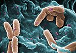 Pseudomonas aeruginosa (bacteria) Pseudomonas aeruginosa (bacteria) | |||||||||||||||||||||||||||
| Method | ELECTRON MICROSCOPY / single particle reconstruction / cryo EM / Resolution: 2.89 Å | |||||||||||||||||||||||||||
 Authors Authors | Jiao, L. / Yihua, H. / Jiao, L. | |||||||||||||||||||||||||||
| Funding support |  China, 1items China, 1items
| |||||||||||||||||||||||||||
 Citation Citation | Journal: Nat Commun / Year: 2026 Title: Structural insights into PSL polysaccharide assembly and export to the cell surface via the Wzx/Wzy-dependent pathway. Authors: Jiao Liu / Yanlong Han / Lanxin Wang / Luyan Z Ma / Dongchun Ni / Yihua Huang / Abstract: Bacterial extracellular polysaccharides play a crucial role in mediating pathogen-host interactions and bacterial fitness via biofilm formation. The Wzx/Wzy-dependent pathway is the most prevalent ...Bacterial extracellular polysaccharides play a crucial role in mediating pathogen-host interactions and bacterial fitness via biofilm formation. The Wzx/Wzy-dependent pathway is the most prevalent and conserved strategy for polysaccharide biosynthesis. Psl (polysaccharide synthesis locus) is a key biofilm matrix polysaccharide in Pseudomonas aeruginosa PAO1, and its biosynthesis machinery is predicted to be a Wzx/Wzy-dependent biosynthesis system. Here, using Psl in PAO1 as a model strain, we determine the cryo-EM structures of the PslD-PslE complex. These structures reveal that PslD-PslE complex forms a continuous, protected conduit across the entire cell envelope. Further structural and functional analyses demonstrate that the polymerase PslJ, is likely localized in the membrane lumen formed by the octameric arrangement of PslE's transmembrane helical pairs. We propose a mechanistic model in which Und-PP-linked pentasaccharide units of Psl access PslJ through side portals in the PslE octamer, shielding the polymerization and translocation processes from degradation by PlsG, a periplasm-localized endoglycosidase. The iterative addition of incoming repeat units to the reducing terminus of the growing polysaccharide chain is hypothesized to drive Psl export through the channel, a mechanism that may be conserved across the Wzx/Wzy-dependent polysaccharide biosynthesis pathways. | |||||||||||||||||||||||||||
| History |
|
- Structure visualization
| Structure viewer | Molecule: MolmilJmol/JSmol |
|---|
- Downloads & links
-Download
| PDBx/mmCIF format | 9v75.cif.gz | 1.2 MB | Display | PDBx/mmCIF format |
|---|---|---|---|---|
| PDB format | pdb9v75.ent.gz | 1007 KB | Display | PDB format |
| PDBx/mmJSON format | 9v75.json.gz | Tree view | PDBx/mmJSON format | |
| Others |  Other downloads Other downloads |
-Validation report
| Arichive directory | https://data.pdbj.org/pub/pdb/validation_reports/v7/9v75ftp://data.pdbj.org/pub/pdb/validation_reports/v7/9v75 | HTTPS FTP |
|---|
-Related structure data
| Related structure data |  64810MC  9v6lC M: map data used to model this data C: citing same article ( |
|---|---|
| Similar structure data |

-Links
 PDBj
PDBj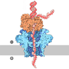
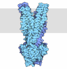
- Assembly
| Deposited unit | 
|
|---|---|
| 1 |
|
-Components
| #1: Protein | Mass: 74543.523 Da / Num. of mol.: 8 Source method: isolated from a genetically manipulated source Source: (gene. exp.) Pseudomonas aeruginosa (bacteria)Gene: ptk, CAZ10_28150, GNQ48_26400, PAERUG_P19_London_7_VIM_2_05_10_00389 Production host:  Escherichia coli (E. coli) Escherichia coli (E. coli)References: UniProt: A0A073A0N3, Transferases; Transferring phosphorus-containing groups; Protein-tyrosine kinases #2: Protein | Mass: 27938.232 Da / Num. of mol.: 8 Source method: isolated from a genetically manipulated source Source: (gene. exp.) Pseudomonas aeruginosa (bacteria)Gene: CAZ10_28145, CSB93_4277, GNQ48_26405, IPC1295_16340, L4V69_19100 Production host: Escherichia coli (E. coli) / References: UniProt: A0A140SD11Has protein modification | Y | |
|---|
-Experimental details
-Experiment
| Experiment | Method: ELECTRON MICROSCOPY |
|---|---|
| EM experiment | Aggregation state: PARTICLE / 3D reconstruction method: single particle reconstruction |
- Sample preparation
| Component | Name: dual complex of Psl transport channel PslD and PslE / Type: COMPLEX / Entity ID: all / Source: RECOMBINANT |
|---|---|
| Molecular weight | Value: 828 MDa / Experimental value: NO |
| Source (natural) | Organism: Pseudomonas aeruginosa (bacteria) |
| Source (recombinant) | Organism: Escherichia coli (E. coli) |
| Buffer solution | pH: 8 |
| Specimen | Embedding applied: NO / Shadowing applied: NO / Staining applied: NO / Vitrification applied: YES |
| Vitrification | Cryogen name: NITROGEN |
- Electron microscopy imaging
| Experimental equipment |  Model: Titan Krios / Image courtesy: FEI Company |
|---|---|
| Microscopy | Model: TFS KRIOS |
| Electron gun | Electron source:  FIELD EMISSION GUN / Accelerating voltage: 300 kV / Illumination mode: FLOOD BEAM FIELD EMISSION GUN / Accelerating voltage: 300 kV / Illumination mode: FLOOD BEAM |
| Electron lens | Mode: BRIGHT FIELD / Nominal magnification: 96000 X / Nominal defocus max: 2000 nm / Nominal defocus min: 1200 nm / Cs: 2.7 mm |
| Specimen holder | Cryogen: NITROGEN |
| Image recording | Electron dose: 60 e/Å2 / Film or detector model: GATAN K3 (6k x 4k) |
- Processing
| EM software |
| |||||||||
|---|---|---|---|---|---|---|---|---|---|---|
| CTF correction | Type: NONE | |||||||||
| 3D reconstruction | Resolution: 2.89 Å / Resolution method: FSC 0.143 CUT-OFF / Num. of particles: 116722 / Symmetry type: POINT |
