 Movie
Movie Controller

[English] 日本語
 Yorodumi
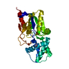
- PDB-9v14: crystal structure of the enzyme-product complex of L-azetidine-2-... -
+ Open data

- Basic information
| Entry | Database: PDB / ID: 9v14 | ||||||
|---|---|---|---|---|---|---|---|
| Title | crystal structure of the enzyme-product complex of L-azetidine-2-carboxylate hydrolase | ||||||
 Components Components | L-azetidine-2-carboxylate hydrolase | ||||||
 Keywords Keywords | HYDROLASE / azetidine-2-carboxylate / alpha/beta hydrolase / dehalogenase-like | ||||||
| Function / homology |  Function and homology information Function and homology informationhydrolase activity, acting on halide bonds, in C-halide compounds / Hydrolases; Acting on carbon-nitrogen bonds, other than peptide bonds / response to toxic substance / periplasmic space Similarity search - Function | ||||||
| Biological species |  Pseudomonas sp. A2C (bacteria) Pseudomonas sp. A2C (bacteria) | ||||||
| Method |  X-RAY DIFFRACTION / SYNCHROTRON / MOLECULAR REPLACEMENT / Resolution: 0.93 Å X-RAY DIFFRACTION / SYNCHROTRON / MOLECULAR REPLACEMENT / Resolution: 0.93 Å | ||||||
 Authors Authors | Toyoda, M. / Mizutani, K. / Mikami, B. / Wackett, L.P. / Esaki, N. / Kurihara, T. | ||||||
| Funding support | 1items
| ||||||
 Citation Citation | Journal: To Be Published Title: Research for the crystal structure of L-azetidine-2-carboxylate hydrolase Authors: Toyoda, M. / Mizutani, K. / Mikami, B. / Wackett, L.P. / Esaki, N. / Kurihara, T. | ||||||
| History |
|
- Structure visualization
| Structure viewer | Molecule: MolmilJmol/JSmol |
|---|
- Downloads & links
-Download
| PDBx/mmCIF format | 9v14.cif.gz | 173.3 KB | Display | PDBx/mmCIF format |
|---|---|---|---|---|
| PDB format | pdb9v14.ent.gz | 136.5 KB | Display | PDB format |
| PDBx/mmJSON format | 9v14.json.gz | Tree view | PDBx/mmJSON format | |
| Others |  Other downloads Other downloads |
-Validation report
| Arichive directory | https://data.pdbj.org/pub/pdb/validation_reports/v1/9v14ftp://data.pdbj.org/pub/pdb/validation_reports/v1/9v14 | HTTPS FTP |
|---|
-Related structure data

-Links
 PDBj
PDBj


- Assembly
| Deposited unit | 
| ||||||||
|---|---|---|---|---|---|---|---|---|---|
| 1 |
| ||||||||
| Unit cell |
|
-Components
-Protein , 1 types, 1 molecules A
| #1: Protein | Mass: 27146.562 Da / Num. of mol.: 1 Source method: isolated from a genetically manipulated source Source: (gene. exp.) Pseudomonas sp. A2C (bacteria) / Production host: |
|---|
-Non-polymers , 7 types, 553 molecules 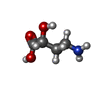


| #2: Chemical | ChemComp-42B /  Mass: 119.119 Da / Num. of mol.: 1 / Source method: obtained synthetically / Formula: C4H9NO3 / Feature type: SUBJECT OF INVESTIGATION Mass: 119.119 Da / Num. of mol.: 1 / Source method: obtained synthetically / Formula: C4H9NO3 / Feature type: SUBJECT OF INVESTIGATION | ||||
|---|---|---|---|---|---|
| #3: Chemical | ChemComp-IMD /  Mass: 69.085 Da / Num. of mol.: 1 / Source method: obtained synthetically / Formula: C3H5N2 Mass: 69.085 Da / Num. of mol.: 1 / Source method: obtained synthetically / Formula: C3H5N2 | ||||
| #4: Chemical | ChemComp-FMT /  Mass: 46.025 Da / Num. of mol.: 1 / Source method: obtained synthetically / Formula: CH2O2 Mass: 46.025 Da / Num. of mol.: 1 / Source method: obtained synthetically / Formula: CH2O2 | ||||
| #5: Chemical | ChemComp-EDO /  Mass: 62.068 Da / Num. of mol.: 1 / Source method: obtained synthetically / Formula: C2H6O2 Mass: 62.068 Da / Num. of mol.: 1 / Source method: obtained synthetically / Formula: C2H6O2 | ||||
| #6: Chemical |  Mass: 106.120 Da / Num. of mol.: 2 / Source method: isolated from a natural source / Formula: C4H10O3 Mass: 106.120 Da / Num. of mol.: 2 / Source method: isolated from a natural source / Formula: C4H10O3#7: Chemical | ChemComp-MG / |  Mass: 24.305 Da / Num. of mol.: 1 / Source method: obtained synthetically / Formula: Mg Mass: 24.305 Da / Num. of mol.: 1 / Source method: obtained synthetically / Formula: Mg#8: Water | ChemComp-HOH / | Mass: 18.015 Da / Num. of mol.: 546 / Source method: isolated from a natural source / Formula: H2O |
-Details
| Has ligand of interest | Y |
|---|---|
| Has protein modification | N |
-Experimental details
-Experiment
| Experiment | Method: X-RAY DIFFRACTION / Number of used crystals: 1 |
|---|
- Sample preparation
| Crystal | Density Matthews: 2.25 Å3/Da / Density % sol: 45.4 % |
|---|---|
| Crystal grow | Temperature: 277 K / Method: vapor diffusion, hanging drop / pH: 8 Details: 22.5% PEG3350, 0.1M imidazole-HCl buffer, 85mM magnesium formate, 50mM sodium azetidine-2-carboxylate |
-Data collection
| Diffraction | Mean temperature: 100 K / Serial crystal experiment: N |
|---|---|
| Diffraction source | Source: SYNCHROTRON / Site: SPring-8  / Beamline: BL26B1 / Wavelength: 0.7 Å / Beamline: BL26B1 / Wavelength: 0.7 Å |
| Detector | Type: RIGAKU RAXIS V / Detector: IMAGE PLATE / Date: Oct 6, 2011 |
| Radiation | Protocol: SINGLE WAVELENGTH / Monochromatic (M) / Laue (L): M / Scattering type: x-ray |
| Radiation wavelength | Wavelength: 0.7 Å / Relative weight: 1 |
| Reflection | Resolution: 0.93→50 Å / Num. obs: 165045 / % possible obs: 100 % / Redundancy: 7 % / CC1/2: 1 / CC star: 1 / Rpim(I) all: 0.019 / Rrim(I) all: 0.05 / Net I/σ(I): 55.2 |
| Reflection shell | Resolution: 0.93→0.95 Å / Redundancy: 6.9 % / Mean I/σ(I) obs: 3.97 / Num. unique obs: 8159 / CC1/2: 0.86 / CC star: 0.962 / Rpim(I) all: 0.311 / Rrim(I) all: 0.821 / % possible all: 100 |
- Processing
| Software |
| ||||||||||||||||||||||||||||||||||||||||||||||||||||||||||||
|---|---|---|---|---|---|---|---|---|---|---|---|---|---|---|---|---|---|---|---|---|---|---|---|---|---|---|---|---|---|---|---|---|---|---|---|---|---|---|---|---|---|---|---|---|---|---|---|---|---|---|---|---|---|---|---|---|---|---|---|---|---|
| Refinement | Method to determine structure: MOLECULAR REPLACEMENT / Resolution: 0.93→10 Å / Cross valid method: FREE R-VALUE
| ||||||||||||||||||||||||||||||||||||||||||||||||||||||||||||
| Refinement step | Cycle: LAST / Resolution: 0.93→10 Å
| ||||||||||||||||||||||||||||||||||||||||||||||||||||||||||||
| LS refinement shell |
|
