 Movie
Movie Controller

[English] 日本語
 Yorodumi
- PDB-8yvw: Crystal structure of D12N mutant of L-azetidine-2-carboxylate hyd... -
+ Open data

- Basic information
| Entry | Database: PDB / ID: 8yvw | ||||||
|---|---|---|---|---|---|---|---|
| Title | Crystal structure of D12N mutant of L-azetidine-2-carboxylate hydrolase | ||||||
 Components Components | (S)-2-haloacid dehalogenase | ||||||
 Keywords Keywords | HYDROLASE / azetidine-2-carboxylate / dehalogenase / alpha/beta hydrolase | ||||||
| Function / homology |  Function and homology information Function and homology informationhydrolase activity, acting on halide bonds, in C-halide compounds / Hydrolases; Acting on carbon-nitrogen bonds, other than peptide bonds / response to toxic substance / periplasmic space Similarity search - Function | ||||||
| Biological species |  Pseudomonas sp. A2C (bacteria) Pseudomonas sp. A2C (bacteria) | ||||||
| Method |  X-RAY DIFFRACTION / SYNCHROTRON / MOLECULAR REPLACEMENT / Resolution: 1.19 Å X-RAY DIFFRACTION / SYNCHROTRON / MOLECULAR REPLACEMENT / Resolution: 1.19 Å | ||||||
 Authors Authors | Toyoda, M. / Mizutani, K. / Mikami, B. / Wackett, L.P. / Esaki, N. / Kurihara, T. | ||||||
| Funding support | 1items
| ||||||
 Citation Citation | Journal: To Be Published Title: Research for the crystal structure of L-azetidine-2-carboxylate hydrolase Authors: Toyoda, M. / Jitsumori, K. / Mikami, B. / Mizutani, K. / Esaki, N. / Wackett, L.P. / Kurihara, T. | ||||||
| History |
|
- Structure visualization
| Structure viewer | Molecule: MolmilJmol/JSmol |
|---|
- Downloads & links
-Download
| PDBx/mmCIF format | 8yvw.cif.gz | 162.3 KB | Display | PDBx/mmCIF format |
|---|---|---|---|---|
| PDB format | pdb8yvw.ent.gz | 105.2 KB | Display | PDB format |
| PDBx/mmJSON format | 8yvw.json.gz | Tree view | PDBx/mmJSON format | |
| Others |  Other downloads Other downloads |
-Validation report
| Arichive directory | https://data.pdbj.org/pub/pdb/validation_reports/yv/8yvwftp://data.pdbj.org/pub/pdb/validation_reports/yv/8yvw | HTTPS FTP |
|---|
-Related structure data
| Related structure data |  8ywoC  9v14C  3smvS S: Starting model for refinement C: citing same article ( |
|---|---|
| Similar structure data |
-Links
 PDBj
PDBj


- Assembly
| Deposited unit | 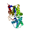
| ||||||||||||
|---|---|---|---|---|---|---|---|---|---|---|---|---|---|
| 1 |
| ||||||||||||
| Unit cell |
|
-Components
| #1: Protein | ( Mass: 27145.578 Da / Num. of mol.: 1 / Mutation: D12N Source method: isolated from a genetically manipulated source Source: (gene. exp.) Pseudomonas sp. A2C (bacteria) / Production host: | ||||||||
|---|---|---|---|---|---|---|---|---|---|
| #2: Chemical |   Mass: 46.025 Da / Num. of mol.: 2 / Source method: obtained synthetically / Formula: CH2O2 / Feature type: SUBJECT OF INVESTIGATION Mass: 46.025 Da / Num. of mol.: 2 / Source method: obtained synthetically / Formula: CH2O2 / Feature type: SUBJECT OF INVESTIGATION#3: Chemical | ChemComp-IMD / |   Mass: 69.085 Da / Num. of mol.: 1 / Source method: obtained synthetically / Formula: C3H5N2 Mass: 69.085 Da / Num. of mol.: 1 / Source method: obtained synthetically / Formula: C3H5N2#4: Chemical | ChemComp-MG / |   Mass: 24.305 Da / Num. of mol.: 1 / Source method: obtained synthetically / Formula: Mg Mass: 24.305 Da / Num. of mol.: 1 / Source method: obtained synthetically / Formula: Mg#5: Water | ChemComp-HOH / |  Mass: 18.015 Da / Num. of mol.: 485 / Source method: isolated from a natural source / Formula: H2O Mass: 18.015 Da / Num. of mol.: 485 / Source method: isolated from a natural source / Formula: H2OHas ligand of interest | Y | |
-Experimental details
-Experiment
| Experiment | Method: X-RAY DIFFRACTION / Number of used crystals: 1 |
|---|
- Sample preparation
| Crystal | Density Matthews: 2.24 Å3/Da / Density % sol: 45.17 % |
|---|---|
| Crystal grow | Temperature: 277 K / Method: vapor diffusion, sitting drop / pH: 8 Details: 25% PEG 3350, 0.1M magnesium formate, 0.1 M imidazole/HCl buffer, pH8.0, 5 mM L-azetidine-2-carboxylate |
-Data collection
| Diffraction | Mean temperature: 100 K / Serial crystal experiment: N |
|---|---|
| Diffraction source | Source: SYNCHROTRON / Site: SPring-8  / Beamline: BL38B1 / Wavelength: 1 Å / Beamline: BL38B1 / Wavelength: 1 Å |
| Detector | Type: RIGAKU JUPITER 210 / Detector: CCD / Date: Mar 8, 2009 |
| Radiation | Protocol: SINGLE WAVELENGTH / Monochromatic (M) / Laue (L): M / Scattering type: x-ray |
| Radiation wavelength | Wavelength: 1 Å / Relative weight: 1 |
| Reflection | Resolution: 1.19→39.52 Å / Num. obs: 76136 / % possible obs: 95.4 % / Redundancy: 9 % / Biso Wilson estimate: 9.54 Å2 / CC1/2: 1 / Rrim(I) all: 0.037 / Net I/σ(I): 37.06 |
| Reflection shell | Resolution: 1.19→1.26 Å / Redundancy: 6.3 % / Mean I/σ(I) obs: 5.26 / Num. unique obs: 10019 / CC1/2: 0.946 / Rrim(I) all: 0.334 / % possible all: 78.7 |
- Processing
| Software |
| ||||||||||||||||||||||||||||||||||||||||||||||||||||||||||||||||||||||||||||||||||||||||||||||||||||||||||||||||||||||||||||||||||||||||||||||||||||||||||||||||||||||||||||||||||||||||||||||||||||
|---|---|---|---|---|---|---|---|---|---|---|---|---|---|---|---|---|---|---|---|---|---|---|---|---|---|---|---|---|---|---|---|---|---|---|---|---|---|---|---|---|---|---|---|---|---|---|---|---|---|---|---|---|---|---|---|---|---|---|---|---|---|---|---|---|---|---|---|---|---|---|---|---|---|---|---|---|---|---|---|---|---|---|---|---|---|---|---|---|---|---|---|---|---|---|---|---|---|---|---|---|---|---|---|---|---|---|---|---|---|---|---|---|---|---|---|---|---|---|---|---|---|---|---|---|---|---|---|---|---|---|---|---|---|---|---|---|---|---|---|---|---|---|---|---|---|---|---|---|---|---|---|---|---|---|---|---|---|---|---|---|---|---|---|---|---|---|---|---|---|---|---|---|---|---|---|---|---|---|---|---|---|---|---|---|---|---|---|---|---|---|---|---|---|---|---|---|---|
| Refinement | Method to determine structure: MOLECULAR REPLACEMENT Starting model: 3SMV Resolution: 1.19→20.38 Å / SU ML: 0.1008 / Cross valid method: FREE R-VALUE / σ(F): 1.34 / Phase error: 12.5303 Stereochemistry target values: GeoStd + Monomer Library + CDL v1.2
| ||||||||||||||||||||||||||||||||||||||||||||||||||||||||||||||||||||||||||||||||||||||||||||||||||||||||||||||||||||||||||||||||||||||||||||||||||||||||||||||||||||||||||||||||||||||||||||||||||||
| Solvent computation | Shrinkage radii: 0.9 Å / VDW probe radii: 1.1 Å / Solvent model: FLAT BULK SOLVENT MODEL | ||||||||||||||||||||||||||||||||||||||||||||||||||||||||||||||||||||||||||||||||||||||||||||||||||||||||||||||||||||||||||||||||||||||||||||||||||||||||||||||||||||||||||||||||||||||||||||||||||||
| Displacement parameters | Biso mean: 13.63 Å2 | ||||||||||||||||||||||||||||||||||||||||||||||||||||||||||||||||||||||||||||||||||||||||||||||||||||||||||||||||||||||||||||||||||||||||||||||||||||||||||||||||||||||||||||||||||||||||||||||||||||
| Refinement step | Cycle: LAST / Resolution: 1.19→20.38 Å
| ||||||||||||||||||||||||||||||||||||||||||||||||||||||||||||||||||||||||||||||||||||||||||||||||||||||||||||||||||||||||||||||||||||||||||||||||||||||||||||||||||||||||||||||||||||||||||||||||||||
| Refine LS restraints |
| ||||||||||||||||||||||||||||||||||||||||||||||||||||||||||||||||||||||||||||||||||||||||||||||||||||||||||||||||||||||||||||||||||||||||||||||||||||||||||||||||||||||||||||||||||||||||||||||||||||
| LS refinement shell |
|
