 Movie
Movie Controller

[English] 日本語
 Yorodumi
- PDB-9uwt: Crystal structure of human galectin-10 produced by cell-free prot... -
+ Open data

- Basic information
| Entry | Database: PDB / ID: 9uwt | ||||||
|---|---|---|---|---|---|---|---|
| Title | Crystal structure of human galectin-10 produced by cell-free protein synthesis in complex with maltose | ||||||
 Components Components | Galectin-10 | ||||||
 Keywords Keywords | SUGAR BINDING PROTEIN / Galectin / CLC | ||||||
| Function / homology |  Function and homology information Function and homology informationregulation of activated T cell proliferation / regulation of T cell cytokine production / T cell apoptotic process / regulation of T cell anergy / carbohydrate binding / extracellular matrix / identical protein binding / cytosol Similarity search - Function | ||||||
| Biological species |  Homo sapiens (human) Homo sapiens (human) | ||||||
| Method |  X-RAY DIFFRACTION / SYNCHROTRON / MOLECULAR REPLACEMENT / Resolution: 2.31 Å X-RAY DIFFRACTION / SYNCHROTRON / MOLECULAR REPLACEMENT / Resolution: 2.31 Å | ||||||
 Authors Authors | Kojima, M. / Ueno, T. / Abe, S. / Hirata, K. | ||||||
| Funding support |  Japan, 1items Japan, 1items
| ||||||
 Citation Citation | Journal: Small Struct / Year: 2025 Title: Cell-Free Protein Crystallization Enables Rapid Structure Determination of Disaccharides and Trisaccharides Using Galectin-10 Crystals. Authors: Kojima, M. / Yao, X. / Abe, S. / Furuta, T. / Hirata, K. / Kobayashi, R. / Suzuki, T. / Ueno, T. | ||||||
| History |
|
- Structure visualization
| Structure viewer | Molecule: MolmilJmol/JSmol |
|---|
- Downloads & links
-Download
| PDBx/mmCIF format | 9uwt.cif.gz | 51.4 KB | Display | PDBx/mmCIF format |
|---|---|---|---|---|
| PDB format | pdb9uwt.ent.gz | 28.9 KB | Display | PDB format |
| PDBx/mmJSON format | 9uwt.json.gz | Tree view | PDBx/mmJSON format | |
| Others |  Other downloads Other downloads |
-Validation report
| Arichive directory | https://data.pdbj.org/pub/pdb/validation_reports/uw/9uwtftp://data.pdbj.org/pub/pdb/validation_reports/uw/9uwt | HTTPS FTP |
|---|
-Related structure data
| Related structure data | 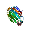 8jaeC  9uwkC  9uwnC  9uwoC  9uwpC  9uwsC  9uwuC C: citing same article ( |
|---|---|
| Similar structure data |
-Links
 PDBj
PDBj


- Assembly
| Deposited unit | 
| ||||||||||||
|---|---|---|---|---|---|---|---|---|---|---|---|---|---|
| 1 | 
| ||||||||||||
| Unit cell |
| ||||||||||||
| Components on special symmetry positions |
|
-Components
| #1: Protein | Mass: 16471.832 Da / Num. of mol.: 1 Source method: isolated from a genetically manipulated source Source: (gene. exp.) Homo sapiens (human) / Gene: CLC, LGALS10, LGALS10A / Production host:  |
|---|---|
| #2: Polysaccharide | alpha-D-glucopyranose-(1-4)-alpha-D-glucopyranose  Source method: isolated from a genetically manipulated source Details: oligosaccharide / References: alpha-maltose |
| #3: Water | ChemComp-HOH /  Mass: 18.015 Da / Num. of mol.: 18 / Source method: isolated from a natural source / Formula: H2O Mass: 18.015 Da / Num. of mol.: 18 / Source method: isolated from a natural source / Formula: H2O |
| Has ligand of interest | Y |
| Has protein modification | N |
-Experimental details
-Experiment
| Experiment | Method: X-RAY DIFFRACTION / Number of used crystals: 1 |
|---|
- Sample preparation
| Crystal | Density Matthews: 2.76 Å3/Da / Density % sol: 55.46 % |
|---|---|
| Crystal grow | Temperature: 293 K / Method: small tubes / Details: cell-free crystallization |
-Data collection
| Diffraction | Mean temperature: 100 K / Serial crystal experiment: N |
|---|---|
| Diffraction source | Source: SYNCHROTRON / Site: SPring-8 / Beamline: BL32XU / Wavelength: 1 Å |
| Detector | Type: DECTRIS PILATUS3 6M / Detector: PIXEL / Date: Jan 20, 2023 |
| Radiation | Protocol: SINGLE WAVELENGTH / Monochromatic (M) / Laue (L): M / Scattering type: x-ray |
| Radiation wavelength | Wavelength: 1 Å / Relative weight: 1 |
| Reflection | Resolution: 2.31→50 Å / Num. obs: 8771 / % possible obs: 96 % / Redundancy: 5.2 % / Biso Wilson estimate: 27.77 Å2 / CC1/2: 0.949 / Net I/σ(I): 4.1 |
| Reflection shell | Resolution: 2.31→2.45 Å / Num. unique obs: 1391 / CC1/2: 0.532 / % possible all: 97.8 |
- Processing
| Software |
| |||||||||||||||||||||||||||||||||||||||||||||||||
|---|---|---|---|---|---|---|---|---|---|---|---|---|---|---|---|---|---|---|---|---|---|---|---|---|---|---|---|---|---|---|---|---|---|---|---|---|---|---|---|---|---|---|---|---|---|---|---|---|---|---|
| Refinement | Method to determine structure: MOLECULAR REPLACEMENT / Resolution: 2.31→35.64 Å / SU ML: 0.3203 / Cross valid method: FREE R-VALUE / σ(F): 1.33 / Phase error: 26.5783 Stereochemistry target values: GeoStd + Monomer Library + CDL v1.2
| |||||||||||||||||||||||||||||||||||||||||||||||||
| Solvent computation | Shrinkage radii: 0.9 Å / VDW probe radii: 1.11 Å / Solvent model: FLAT BULK SOLVENT MODEL | |||||||||||||||||||||||||||||||||||||||||||||||||
| Displacement parameters | Biso mean: 25.82 Å2 | |||||||||||||||||||||||||||||||||||||||||||||||||
| Refinement step | Cycle: LAST / Resolution: 2.31→35.64 Å
| |||||||||||||||||||||||||||||||||||||||||||||||||
| Refine LS restraints |
| |||||||||||||||||||||||||||||||||||||||||||||||||
| LS refinement shell |
|
