 Movie
Movie Controller

[English] 日本語
 Yorodumi
- PDB-9udl: Cocrystal structure of human cytosolic phenylalanyl-tRNA syntheta... -
+ Open data

- Basic information
| Entry | Database: PDB / ID: 9udl | ||||||
|---|---|---|---|---|---|---|---|
| Title | Cocrystal structure of human cytosolic phenylalanyl-tRNA synthetase and an inhibitor | ||||||
 Components Components |
| ||||||
 Keywords Keywords | LIGASE / TRNA ligase / Inhibitor / Complex | ||||||
| Function / homology |  Function and homology information Function and homology informationphenylalanine-tRNA ligase complex / phenylalanine-tRNA ligase / phenylalanyl-tRNA aminoacylation / phenylalanine-tRNA ligase activity / Cytosolic tRNA aminoacylation / protein heterotetramerization / tRNA binding / translation / magnesium ion binding / RNA binding ...phenylalanine-tRNA ligase complex / phenylalanine-tRNA ligase / phenylalanyl-tRNA aminoacylation / phenylalanine-tRNA ligase activity / Cytosolic tRNA aminoacylation / protein heterotetramerization / tRNA binding / translation / magnesium ion binding / RNA binding / ATP binding / membrane / cytoplasm / cytosol Similarity search - Function | ||||||
| Biological species |  Homo sapiens (human) Homo sapiens (human) | ||||||
| Method |  X-RAY DIFFRACTION / SYNCHROTRON / MOLECULAR REPLACEMENT / Resolution: 3.4 Å X-RAY DIFFRACTION / SYNCHROTRON / MOLECULAR REPLACEMENT / Resolution: 3.4 Å | ||||||
 Authors Authors | Qiao, H. / Hei, Z. / Fang, P. | ||||||
| Funding support | 1items
| ||||||
 Citation Citation | Journal: To Be Published Title: Cocrystal structure of human cytosolic phenylalanyl-tRNA synthetase and an inhibitor Authors: Qiao, H. / Hei, Z. / Fang, P. | ||||||
| History |
|
- Structure visualization
| Structure viewer | Molecule: MolmilJmol/JSmol |
|---|
- Downloads & links
-Download
| PDBx/mmCIF format | 9udl.cif.gz | 357.6 KB | Display | PDBx/mmCIF format |
|---|---|---|---|---|
| PDB format | pdb9udl.ent.gz | 291.5 KB | Display | PDB format |
| PDBx/mmJSON format | 9udl.json.gz | Tree view | PDBx/mmJSON format | |
| Others |  Other downloads Other downloads |
-Validation report
| Arichive directory | https://data.pdbj.org/pub/pdb/validation_reports/ud/9udlftp://data.pdbj.org/pub/pdb/validation_reports/ud/9udl | HTTPS FTP |
|---|
-Related structure data
| Related structure data |  9udmC 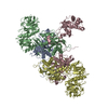 3l4gS S: Starting model for refinement C: citing same article ( |
|---|---|
| Similar structure data |
-Links
 PDBj
PDBj

- Assembly
| Deposited unit | 
| ||||||||
|---|---|---|---|---|---|---|---|---|---|
| 1 | 
| ||||||||
| Unit cell |
|
-Components
| #1: Protein | Mass: 36433.434 Da / Num. of mol.: 1 Source method: isolated from a genetically manipulated source Source: (gene. exp.) Homo sapiens (human) / Gene: FARSA, FARS, FARSL, FARSLA / Production host:  |
|---|---|
| #2: Protein | Mass: 66214.484 Da / Num. of mol.: 1 Source method: isolated from a genetically manipulated source Source: (gene. exp.) Homo sapiens (human) / Gene: FARSB, FARSLB, FRSB, HSPC173 / Production host: |
| #3: Chemical | ChemComp-LCF / [  Mass: 379.879 Da / Num. of mol.: 1 / Source method: obtained synthetically / Formula: C23H22ClNO2 / Feature type: SUBJECT OF INVESTIGATION / Comment: antiinflammatory, inhibitor*YM Mass: 379.879 Da / Num. of mol.: 1 / Source method: obtained synthetically / Formula: C23H22ClNO2 / Feature type: SUBJECT OF INVESTIGATION / Comment: antiinflammatory, inhibitor*YM |
| Has ligand of interest | Y |
| Has protein modification | N |
-Experimental details
-Experiment
| Experiment | Method: X-RAY DIFFRACTION / Number of used crystals: 1 |
|---|
- Sample preparation
| Crystal | Density Matthews: 4.08 Å3/Da / Density % sol: 69.83 % |
|---|---|
| Crystal grow | Temperature: 291 K / Method: vapor diffusion, sitting drop / pH: 8.5 Details: 1.2 M sodium formate, 0.05 M tris pH 8.5, 8% v/v PEG 500 MME and 8% w/v PEG 20000. |
-Data collection
| Diffraction | Mean temperature: 100 K / Serial crystal experiment: N |
|---|---|
| Diffraction source | Source: SYNCHROTRON / Site: SSRF  / Beamline: BL02U1 / Wavelength: 0.97918 Å / Beamline: BL02U1 / Wavelength: 0.97918 Å |
| Detector | Type: DECTRIS EIGER X 9M / Detector: PIXEL / Date: Sep 29, 2023 |
| Radiation | Protocol: SINGLE WAVELENGTH / Monochromatic (M) / Laue (L): M / Scattering type: x-ray |
| Radiation wavelength | Wavelength: 0.97918 Å / Relative weight: 1 |
| Reflection | Resolution: 3.4→48.9 Å / Num. obs: 24301 / % possible obs: 100 % / Redundancy: 12.9 % / CC1/2: 0.999 / Rmerge(I) obs: 0.163 / Rpim(I) all: 0.047 / Rrim(I) all: 0.17 / Χ2: 1.1 / Net I/σ(I): 13.6 / Num. measured all: 314175 |
| Reflection shell | Resolution: 3.4→3.63 Å / % possible obs: 100 % / Redundancy: 13.8 % / Rmerge(I) obs: 2.82 / Num. measured all: 59335 / Num. unique obs: 4314 / CC1/2: 0.57 / Rpim(I) all: 0.787 / Rrim(I) all: 2.929 / Χ2: 0.84 / Net I/σ(I) obs: 1 |
- Processing
| Software |
| ||||||||||||||||||||||||||||||||||||||||||||||||||||||||||||||||||||||
|---|---|---|---|---|---|---|---|---|---|---|---|---|---|---|---|---|---|---|---|---|---|---|---|---|---|---|---|---|---|---|---|---|---|---|---|---|---|---|---|---|---|---|---|---|---|---|---|---|---|---|---|---|---|---|---|---|---|---|---|---|---|---|---|---|---|---|---|---|---|---|---|
| Refinement | Method to determine structure: MOLECULAR REPLACEMENT Starting model: 3L4G Resolution: 3.4→45.35 Å / SU ML: 0.58 / Cross valid method: FREE R-VALUE / σ(F): 1.33 / Phase error: 29.63 / Stereochemistry target values: ML
| ||||||||||||||||||||||||||||||||||||||||||||||||||||||||||||||||||||||
| Solvent computation | Shrinkage radii: 0.9 Å / VDW probe radii: 1.1 Å / Solvent model: FLAT BULK SOLVENT MODEL | ||||||||||||||||||||||||||||||||||||||||||||||||||||||||||||||||||||||
| Refinement step | Cycle: LAST / Resolution: 3.4→45.35 Å
| ||||||||||||||||||||||||||||||||||||||||||||||||||||||||||||||||||||||
| Refine LS restraints |
| ||||||||||||||||||||||||||||||||||||||||||||||||||||||||||||||||||||||
| LS refinement shell |
| ||||||||||||||||||||||||||||||||||||||||||||||||||||||||||||||||||||||
| Refinement TLS params. | Method: refined / Origin x: 34.8032 Å / Origin y: -40.8683 Å / Origin z: 18.5964 Å
| ||||||||||||||||||||||||||||||||||||||||||||||||||||||||||||||||||||||
| Refinement TLS group | Selection details: all |
