 Movie
Movie Controller


+ Open data

- Basic information
| Entry | Database: PDB / ID: 9u9h | |||||||||||||||||||||
|---|---|---|---|---|---|---|---|---|---|---|---|---|---|---|---|---|---|---|---|---|---|---|
| Title | Surface Tubular Element of Vaccinia Virus | |||||||||||||||||||||
 Components Components |
| |||||||||||||||||||||
 Keywords Keywords | VIRAL PROTEIN / poxvirus / membrane protein | |||||||||||||||||||||
| Function / homology |  Function and homology information Function and homology informationhelicase activity / DNA-templated transcription termination / hydrolase activity / viral envelope / virion membrane / DNA binding / ATP binding / membrane Similarity search - Function | |||||||||||||||||||||
| Biological species |  Vaccinia virus Vaccinia virus | |||||||||||||||||||||
| Method | ELECTRON MICROSCOPY / helical reconstruction / cryo EM / Resolution: 3.23 Å | |||||||||||||||||||||
 Authors Authors | Yu, F. / Jin, G. / Liu, Y. / Sun, Z. / Lou, Z. | |||||||||||||||||||||
| Funding support |  China, 2items China, 2items
| |||||||||||||||||||||
 Citation Citation | Journal: mBio / Year: 2026 Title: Architecture of surface tubular element of poxvirus. Authors: Fengxi Yu / Ge Jin / Yixiao Liu / Zhenyu Liu / Jingxuan Yao / Junbo Wang / Daoxin Xie / Zihe Rao / Liming Yan / Yan Zhang / Zixian Sun / Zhiyong Lou / Abstract: Poxviruses are large enveloped DNA viruses that cause severe human infectious diseases. The mature virion of poxvirus is covered with dense surface tubular elements (STEs), which play a role in ...Poxviruses are large enveloped DNA viruses that cause severe human infectious diseases. The mature virion of poxvirus is covered with dense surface tubular elements (STEs), which play a role in assembly progress of mature virions (MVs) and inhibit host cell protein synthesis. However, the composition and assembly of STEs remain unclear. Cryo-electron microscopy (cryo-EM) has proven to be a powerful technique for determining the structure of proteins from complex biological samples. By integrating high-resolution cryo-EM maps with mass spectrometry, we reveal that STEs are helically assembled from two transmembrane proteins, A14 and A17, which bind to phospholipid molecules and form the tubular scaffold along the poxviral membrane. Extensive intermolecular interactions, including A14 dimers and A14-A17 complexes, drive the remarkable structural stability of STEs. Structural analysis further emphasizes the reticulon-like properties of A17, which promote membrane curvature and stabilize the tubular architecture. These results provide novel insights into the STE assembly, morphogenesis, and surface organization of poxviruses, offering valuable information for the development of vaccines and antiviral strategies against poxvirus infections.IMPORTANCESurface tubular elements (STEs) are critical components of poxvirus mature virions and play a role in suppressing host cell protein synthesis. In this study, we isolated and purified STEs from native poxvirus virions and subsequently determined their core composition and high-resolution architecture. We identified that STE is mainly composed of membrane proteins A14 and A17, along with phospholipid molecules. Within the repeat structural unit of STE, A14 proteins form two homodimers within the repeating unit, with A17 monomers flanking either side. Phospholipid molecules are distributed within the A14-A14 and A14-A17 interfaces. Our study not only revealed the molecular structures of A14 and A17 but also further emphasized that the reticulon-like and highly oligomerized characteristics of A17 provide membrane curvature, while the A14-A17-phospholipid network stabilizes the tubular structure. We proposed a hypothetical model that A17 drives changes in viral membrane curvature during maturation. These findings enhance our understanding of poxvirus biology and may guide therapeutic strategies against poxvirus infections. | |||||||||||||||||||||
| History |
|
- Structure visualization
| Structure viewer | Molecule: MolmilJmol/JSmol |
|---|
- Downloads & links
-Download
| PDBx/mmCIF format | 9u9h.cif.gz | 150 KB | Display | PDBx/mmCIF format |
|---|---|---|---|---|
| PDB format | pdb9u9h.ent.gz | 119.2 KB | Display | PDB format |
| PDBx/mmJSON format | 9u9h.json.gz | Tree view | PDBx/mmJSON format | |
| Others |  Other downloads Other downloads |
-Validation report
| Arichive directory | https://data.pdbj.org/pub/pdb/validation_reports/u9/9u9hftp://data.pdbj.org/pub/pdb/validation_reports/u9/9u9h | HTTPS FTP |
|---|
-Related structure data
| Related structure data |  63964MC M: map data used to model this data C: citing same article ( |
|---|---|
| Similar structure data |
-Links
 PDBj
PDBj

- Assembly
| Deposited unit | 
|
|---|---|
| 1 |
|
-Components
| #1: Protein | Mass: 10003.072 Da / Num. of mol.: 4 / Source method: isolated from a natural source Details: Sequence reference for source organism Vaccinia virus (strain Tian Tan) is not available in UniProt at the time of biocuration. Current sequence reference is from UniProt id P20991. Source: (natural) Vaccinia virus (strain Tian Tan) / References: UniProt: P20991#2: Protein | Mass: 19027.289 Da / Num. of mol.: 4 / Source method: isolated from a natural source Details: Sequence reference for source organism Vaccinia virus (strain Tian Tan) is not available in UniProt at the time of biocuration. Current sequence reference is from UniProt id P68592. Source: (natural) Vaccinia virus (strain Tian Tan) / References: UniProt: P68592#3: Chemical | ChemComp-PX4 / 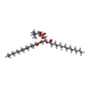  Mass: 678.940 Da / Num. of mol.: 9 / Source method: obtained synthetically / Formula: C36H73NO8P / Feature type: SUBJECT OF INVESTIGATION / Comment: DMPC, phospholipid*YM Mass: 678.940 Da / Num. of mol.: 9 / Source method: obtained synthetically / Formula: C36H73NO8P / Feature type: SUBJECT OF INVESTIGATION / Comment: DMPC, phospholipid*YMHas ligand of interest | Y | Has protein modification | N | |
|---|
-Experimental details
-Experiment
| Experiment | Method: ELECTRON MICROSCOPY |
|---|---|
| EM experiment | Aggregation state: FILAMENT / 3D reconstruction method: helical reconstruction |
- Sample preparation
| Component | Name: Surface Tubular Element of Vaccinia Virus / Type: COMPLEX / Entity ID: #1-#2 / Source: NATURAL |
|---|---|
| Source (natural) | Organism: Vaccinia virus / Strain: Non-replicating vaccinia virus TianTan Strain |
| Buffer solution | pH: 8 |
| Specimen | Embedding applied: NO / Shadowing applied: NO / Staining applied: NO / Vitrification applied: YES |
| Vitrification | Cryogen name: ETHANE |
- Electron microscopy imaging
| Experimental equipment |  Model: Titan Krios / Image courtesy: FEI Company |
|---|---|
| Microscopy | Model: TFS KRIOS |
| Electron gun | Electron source:  FIELD EMISSION GUN / Accelerating voltage: 300 kV / Illumination mode: FLOOD BEAM FIELD EMISSION GUN / Accelerating voltage: 300 kV / Illumination mode: FLOOD BEAM |
| Electron lens | Mode: BRIGHT FIELD / Nominal defocus max: 4000 nm / Nominal defocus min: 1000 nm |
| Image recording | Electron dose: 60 e/Å2 / Film or detector model: GATAN K2 BASE (4k x 4k) |
- Processing
| CTF correction | Type: PHASE FLIPPING AND AMPLITUDE CORRECTION |
|---|---|
| Helical symmerty | Angular rotation/subunit: -50 ° / Axial rise/subunit: 26.58 Å / Axial symmetry: C1 |
| 3D reconstruction | Resolution: 3.23 Å / Resolution method: FSC 0.143 CUT-OFF / Num. of particles: 477012 / Algorithm: FOURIER SPACE / Symmetry type: HELICAL |
