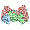
[English] 日本語
Yorodumi
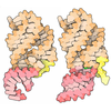
- PDB-9tjv: Ternary complex of E. coli leucyl-tRNA synthetase mutant W177A, t... -

+
Open data


ID or keywords:

Loading...

-
Basic information

Entry
Database: PDB / ID: 9tjv
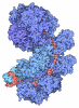
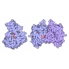
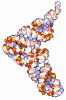
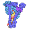
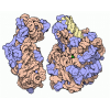
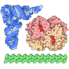
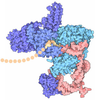
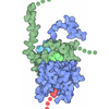
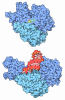
TitleTernary complex of E. coli leucyl-tRNA synthetase mutant W177A, tRNA(leu) and the leucyl adenylate analog LeuAMS in the aminoacyl transfer state
Components
  • Leucine--tRNA ligase
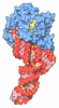
  • tRNA(leu)
KeywordsRNA BINDING PROTEIN / Leucine tRNA ligase Antimicrobial target tRNA aminoacylation for protein translation
Function / homology
Function and homology information


leucine-tRNA ligase / leucine-tRNA ligase activity / leucyl-tRNA aminoacylation / aminoacyl-tRNA deacylase activity / ATP binding / cytosol
Similarity search - Function
Leucyl-tRNA synthetase, editing domain / Leucyl-tRNA synthetase, editing domain / Leucine-tRNA ligase / Aminoacyl-tRNA synthetase, class Ia / tRNA synthetases class I (I, L, M and V) / Valyl/Leucyl/Isoleucyl-tRNA synthetase, editing domain / Methionyl/Valyl/Leucyl/Isoleucyl-tRNA synthetase, anticodon-binding / Anticodon-binding domain of tRNA ligase / Methionyl/Leucyl tRNA synthetase / tRNA synthetases class I (M) ...Leucyl-tRNA synthetase, editing domain / Leucyl-tRNA synthetase, editing domain / Leucine-tRNA ligase / Aminoacyl-tRNA synthetase, class Ia / tRNA synthetases class I (I, L, M and V) / Valyl/Leucyl/Isoleucyl-tRNA synthetase, editing domain / Methionyl/Valyl/Leucyl/Isoleucyl-tRNA synthetase, anticodon-binding / Anticodon-binding domain of tRNA ligase / Methionyl/Leucyl tRNA synthetase / tRNA synthetases class I (M) / Aminoacyl-tRNA synthetase, class Ia, anticodon-binding / Aminoacyl-tRNA synthetase, class I, conserved site / Aminoacyl-transfer RNA synthetases class-I signature. / Rossmann-like alpha/beta/alpha sandwich fold
Similarity search - Domain/homology
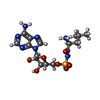
5'-O-(L-leucylsulfamoyl)adenosine / : / RNA / RNA (> 10) / Leucine--tRNA ligase
Similarity search - Component
Biological speciesEscherichia coli (E. coli)
MethodX-RAY DIFFRACTION / SYNCHROTRON / MOLECULAR REPLACEMENT / Resolution: 2.277 Å
AuthorsHoffmann, G. / Palencia, A.
Funding support France, European Union, 4items
OrganizationGrant numberCountry
Grenoble Instruct-ERIC Center (ISBG)27604 France
iNEXT-Discovery45625European Union
Agence Nationale de la Recherche (ANR)ANR-20-AMRB-0003 France
Agence Nationale de la Recherche (ANR)ANR-22-CE44-0040 France
CitationJournal: Nucleic Acids Res. / Year: 2026
Title: The Zn Domain Acts as a Dynamic Switch Coordinating Multiple-Step Aminoacylation in Bacterial Leucyl-tRNA Synthetase
Authors: Hoffmann, G. / Dulic, M. / Gruic-Sovulj, I. / Palencia, A.
History
DepositionDec 8, 2025Deposition site: PDBE / Processing site: PDBE
Revision 1.0Aug 5, 2026Provider: repository / Type: Initial release

-
Structure visualization

Structure viewerMolecule:
MolmilJmol/JSmol

Downloads & links

-
Assembly

Deposited unit
A: Leucine--tRNA ligase
B: tRNA(leu)
C: Leucine--tRNA ligase
D: tRNA(leu)
hetero molecules


Theoretical massNumber of molelcules
Total (without water)255,8758
Polymers254,9074
Non-polymers9684
Water12,520695
1
A: Leucine--tRNA ligase
B: tRNA(leu)
hetero molecules


Theoretical massNumber of molelcules
Total (without water)127,9625
Polymers127,4542
Non-polymers5083
Water362
TypeNameSymmetry operationNumber
identity operation1_555x,y,z1
Buried area5770 Å2
ΔGint-63 kcal/mol
Surface area46060 Å2
MethodPISA
2
C: Leucine--tRNA ligase
D: tRNA(leu)
hetero molecules


Theoretical massNumber of molelcules
Total (without water)127,9133
Polymers127,4542
Non-polymers4591
Water362
TypeNameSymmetry operationNumber
identity operation1_555x,y,z1
Buried area5530 Å2
ΔGint-42 kcal/mol
Surface area46690 Å2
MethodPISA
Unit cell
Length a, b, c (Å)158.009, 68.312, 226.276
Angle α, β, γ (deg.)90, 104.44, 90
Int Tables number5
Space group name H-MC121

-
Components

#1: Protein Leucine--tRNA ligase / Leucyl-tRNA synthetase / LeuRS


Mass: 99400.883 Da / Num. of mol.: 2 / Mutation: W177A
Source method: isolated from a genetically manipulated source
Details: E. coli Leucyl tRNA synthetase with W177A mutation / Source: (gene. exp.) Escherichia coli (E. coli) / Gene: leuS, BWG_0513 / Production host: Escherichia coli (E. coli) / References: UniProt: C4ZWC9, leucine-tRNA ligase
#2: RNA chain tRNA(leu)


Mass: 28052.652 Da / Num. of mol.: 2
Source method: isolated from a genetically manipulated source
Details: E. coli tRNA(Leu) isoacceptor UAA / Source: (gene. exp.) Escherichia coli (E. coli)
Production host: in vitro transcription vector pT7-Fluc(deltai) (others)
References: GenBank: 1231762938
#3: Chemical ChemComp-LSS / 5'-O-(L-leucylsulfamoyl)adenosine / 5-O-N-LEUCYL-SULFAMOYLADENOSINE


Mass: 459.477 Da / Num. of mol.: 2 / Source method: obtained synthetically / Formula: C16H25N7O7S / Feature type: SUBJECT OF INVESTIGATION
#4: Chemical ChemComp-MG / MAGNESIUM ION


Mass: 24.305 Da / Num. of mol.: 2 / Source method: obtained synthetically / Formula: Mg
#5: Water ChemComp-HOH / water


Mass: 18.015 Da / Num. of mol.: 695 / Source method: isolated from a natural source / Formula: H2O
Has ligand of interestY
Has protein modificationN

-
Experimental details

-
Experiment

ExperimentMethod: X-RAY DIFFRACTION / Number of used crystals: 1

-
Sample preparation

CrystalDensity Matthews: 2.32 Å3/Da / Density % sol: 46.98 %
Crystal growTemperature: 293 K / Method: vapor diffusion, hanging drop / pH: 5.5
Details: 0.1M BisTris pH5.5, 0.2M Ammonium Acetate, 23-26% PEG3350

-
Data collection

DiffractionMean temperature: 100 K / Serial crystal experiment: N
Diffraction sourceSource: SYNCHROTRON / Site: ESRF / Beamline: ID30B / Wavelength: 0.87313 Å
DetectorType: DECTRIS EIGER2 S 9M / Detector: PIXEL / Date: Mar 7, 2024
RadiationProtocol: SINGLE WAVELENGTH / Monochromatic (M) / Laue (L): M / Scattering type: x-ray
Radiation wavelengthWavelength: 0.87313 Å / Relative weight: 1
ReflectionResolution: 2.28→219.132 Å / Num. obs: 84189 / % possible obs: 92 % / Redundancy: 3.3 % / Biso Wilson estimate: 40.1 Å2 / CC1/2: 0.992 / Rmerge(I) obs: 0.127 / Rpim(I) all: 0.083 / Rrim(I) all: 0.152 / Net I/σ(I): 5.2
Reflection shellResolution: 2.28→2.49 Å / Redundancy: 3.4 % / Rmerge(I) obs: 0.773 / Mean I/σ(I) obs: 1.5 / Num. unique obs: 4209 / CC1/2: 0.591 / Rpim(I) all: 0.486 / Rrim(I) all: 0.915 / % possible all: 43.9

-
Processing

Software
NameVersionClassification
BUSTER2.10.4refinement
autoPROC20240123data reduction
STARANISOdata scaling
PHASER2.8.3phasing
RefinementMethod to determine structure: MOLECULAR REPLACEMENT / Resolution: 2.277→219.13 Å / Cor.coef. Fo:Fc: 0.94 / Cor.coef. Fo:Fc free: 0.924 / SU R Cruickshank DPI: 0.537 / Cross valid method: THROUGHOUT / SU R Blow DPI: 0.574 / SU Rfree Blow DPI: 0.249 / SU Rfree Cruickshank DPI: 0.249
RfactorNum. reflection% reflectionSelection details
Rfree0.2224 4265 -RANDOM
Rwork0.1938 79924 --
obs0.1953 84189 78.3 %-
Displacement parametersBiso mean: 67.43 Å2
Baniso -1Baniso -2Baniso -3
1-0.31 Å20 Å20.5856 Å2
2---1.5616 Å20 Å2
3---1.2517 Å2
Refine analyzeLuzzati coordinate error obs: 0.291 Å
Refinement stepCycle: LAST / Resolution: 2.277→219.13 Å
ProteinNucleic acidLigandSolventTotal
Num. atoms13534 3307 64 695 17600
Refine LS restraints
Refine-IDTypeDev idealNumberRestraint functionWeight
X-RAY DIFFRACTIONt_bond_d0.00717607HARMONIC2
X-RAY DIFFRACTIONt_angle_deg0.8124649HARMONIC2
X-RAY DIFFRACTIONt_dihedral_angle_d5572SINUSOIDAL2
X-RAY DIFFRACTIONt_gen_planes2529HARMONIC5
X-RAY DIFFRACTIONt_it17607HARMONIC10
X-RAY DIFFRACTIONt_chiral_improper_torsion2393SEMIHARMONIC5
X-RAY DIFFRACTIONt_ideal_dist_contact12258SEMIHARMONIC4
X-RAY DIFFRACTIONt_omega_torsion2.9
X-RAY DIFFRACTIONt_other_torsion16.85
LS refinement shellResolution: 2.28→2.43 Å
RfactorNum. reflection% reflection
Rfree0.2928 88 -
Rwork0.2546 1596 -
obs0.2566 1684 9 %
Refinement TLS params.

Refine-ID: X-RAY DIFFRACTION

IDL112)L122)L132)L222)L232)L332)S11 (Å °)S12 (Å °)S13 (Å °)S21 (Å °)S22 (Å °)S23 (Å °)S31 (Å °)S32 (Å °)S33 (Å °)T112)T122)T132)T222)T232)T332)Origin x (Å)Origin y (Å)Origin z (Å)
11.24310.2550.45781.08490.05610.2374-0.1177-0.15790.14260.12290.06230.2551-0.0274-0.05530.0554-0.08130.00040.0769-0.03960.00720.0432-57.83879.1478-85.2114
20.20030.04840.04190.3874-0.19870.09510.0112-0.05820.01720.057-0.01040.0103-0.01480.0036-0.0008-0.0165-0.01210.0317-0.0689-0.01250.0288-31.70398.9699-84.116
33.93190.4503-0.54320.4888-0.82740.7826-0.002-0.8143-0.5677-0.292-0.0359-0.0890.4202-0.05440.03790.24190.0842-0.03310.09520.2775-0.4606-69.603313.487-27.515
41.04410.64070.40290.4035-0.66531.11230.1429-0.27450.0991-0.0692-0.14690.010.13230.08220.0040.08860.1785-0.00460.13180.0354-0.4121-52.22732.1998-29.2948
Refinement TLS group
IDRefine-IDRefine TLS-IDSelection detailsAuth asym-IDAuth seq-ID
1X-RAY DIFFRACTION1{ B|* }B1 - 76
2X-RAY DIFFRACTION2{ A|* }A0 - 860
3X-RAY DIFFRACTION2{ A|* }A901
4X-RAY DIFFRACTION3{ D|* }D1 - 76
5X-RAY DIFFRACTION4{ C|* }C1 - 860
6X-RAY DIFFRACTION4{ C|* }C901

+
About Yorodumi

-
News

-
Feb 9, 2022. New format data for meta-information of EMDB entries

New format data for meta-information of EMDB entries

  • Version 3 of the EMDB header file is now the official format.
  • The previous official version 1.9 will be removed from the archive.

Related info.:EMDB header

External links:wwPDB to switch to version 3 of the EMDB data model

-
Aug 12, 2020. Covid-19 info

Covid-19 info

URL: https://pdbj.org/emnavi/covid19.php

New page: Covid-19 featured information page in EM Navigator.

Related info.:Covid-19 info / Mar 5, 2020. Novel coronavirus structure data

+
Mar 5, 2020. Novel coronavirus structure data

Novel coronavirus structure data

Related info.:Yorodumi Speices / Aug 12, 2020. Covid-19 info

External links:COVID-19 featured content - PDBj / Molecule of the Month (242):Coronavirus Proteases

+
Jan 31, 2019. EMDB accession codes are about to change! (news from PDBe EMDB page)

EMDB accession codes are about to change! (news from PDBe EMDB page)

  • The allocation of 4 digits for EMDB accession codes will soon come to an end. Whilst these codes will remain in use, new EMDB accession codes will include an additional digit and will expand incrementally as the available range of codes is exhausted. The current 4-digit format prefixed with “EMD-” (i.e. EMD-XXXX) will advance to a 5-digit format (i.e. EMD-XXXXX), and so on. It is currently estimated that the 4-digit codes will be depleted around Spring 2019, at which point the 5-digit format will come into force.
  • The EM Navigator/Yorodumi systems omit the EMD- prefix.

Related info.:Q: What is EMD? / ID/Accession-code notation in Yorodumi/EM Navigator

External links:EMDB Accession Codes are Changing Soon! / Contact to PDBj

+
Jul 12, 2017. Major update of PDB

Major update of PDB

  • wwPDB released updated PDB data conforming to the new PDBx/mmCIF dictionary.
  • This is a major update changing the version number from 4 to 5, and with Remediation, in which all the entries are updated.
  • In this update, many items about electron microscopy experimental information are reorganized (e.g. em_software).
  • Now, EM Navigator and Yorodumi are based on the updated data.

External links:wwPDB Remediation / Enriched Model Files Conforming to OneDep Data Standards Now Available in the PDB FTP Archive

-
Yorodumi

Thousand views of thousand structures

  • Yorodumi is a browser for structure data from EMDB, PDB, SASBDB, etc.
  • This page is also the successor to EM Navigator detail page, and also detail information page/front-end page for Omokage search.
  • The word "yorodu" (or yorozu) is an old Japanese word meaning "ten thousand". "mi" (miru) is to see.

Related info.:EMDB / PDB / SASBDB / Comparison of 3 databanks / Yorodumi Search / Aug 31, 2016. New EM Navigator & Yorodumi / Yorodumi Papers / Jmol/JSmol / Function and homology information / Changes in new EM Navigator and Yorodumi

Read more