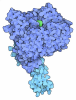
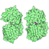
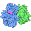
[English] 日本語
Yorodumi
- PDB-9tg7: Crystal structure of beta-TrCP bound by monophosphorylated WEE1 d... -

+
Open data


ID or keywords:

Loading...

-
Basic information

Entry
Database: PDB / ID: 9tg7
TitleCrystal structure of beta-TrCP bound by monophosphorylated WEE1 degron peptide
Components
  • F-box/WD repeat-containing protein 1A
  • Wee1-like protein kinase
KeywordsTRANSFERASE / E3 ligase / phosphodegron / beta-TrCP / WEE1
Function / homology
Function and homology information


protein phosphorylated amino acid binding / regulation of cell cycle process / negative regulation of G2/M transition of mitotic cell cycle / G2/M DNA replication checkpoint / positive regulation of circadian rhythm / Polo-like kinase mediated events / ubiquitin ligase activator activity / regulation of canonical Wnt signaling pathway / protein dephosphorylation / negative regulation of G1/S transition of mitotic cell cycle ...protein phosphorylated amino acid binding / regulation of cell cycle process / negative regulation of G2/M transition of mitotic cell cycle / G2/M DNA replication checkpoint / positive regulation of circadian rhythm / Polo-like kinase mediated events / ubiquitin ligase activator activity / regulation of canonical Wnt signaling pathway / protein dephosphorylation / negative regulation of G1/S transition of mitotic cell cycle / non-canonical NF-kappaB signal transduction / SCF ubiquitin ligase complex / SCF-dependent proteasomal ubiquitin-dependent protein catabolic process / ligase activity / Prolactin receptor signaling / positive regulation of proteolysis / negative regulation of T cell receptor signaling pathway / Cyclin E associated events during G1/S transition / Cyclin A:Cdk2-associated events at S phase entry / GSK3B-mediated proteasomal degradation of PD-L1(CD274) / Chk1/Chk2(Cds1) mediated inactivation of Cyclin B:Cdk1 complex / Cyclin A/B1/B2 associated events during G2/M transition / ubiquitin-like ligase-substrate adaptor activity / protein K48-linked ubiquitination / positive regulation of DNA replication / negative regulation of smoothened signaling pathway / MAP3K8 (TPL2)-dependent MAPK1/3 activation / non-specific protein-tyrosine kinase / non-membrane spanning protein tyrosine kinase activity / Deactivation of the beta-catenin transactivating complex / SCF-beta-TrCP mediated degradation of Emi1 / NIK-->noncanonical NF-kB signaling / Vpu mediated degradation of CD4 / protein destabilization / Dectin-1 mediated noncanonical NF-kB signaling / Degradation of CRY and PER proteins / Activation of NF-kappaB in B cells / Degradation of GLI1 by the proteasome / GSK3B and BTRC:CUL1-mediated-degradation of NFE2L2 / regulation of circadian rhythm / beta-catenin binding / Degradation of GLI2 by the proteasome / GLI3 is processed to GLI3R by the proteasome / Ubiquitin-Mediated Degradation of Phosphorylated Cdc25A / Wnt signaling pathway / G2/M transition of mitotic cell cycle / Degradation of beta-catenin by the destruction complex / CLEC7A (Dectin-1) signaling / FCERI mediated NF-kB activation / protein polyubiquitination / Interleukin-1 signaling / ubiquitin protein ligase activity / Regulation of PLK1 Activity at G2/M Transition / rhythmic process / Downstream TCR signaling / Antigen processing: Ubiquitination & Proteasome degradation / Factors involved in megakaryocyte development and platelet production / Neddylation / protein tyrosine kinase activity / ubiquitin-dependent protein catabolic process / proteasome-mediated ubiquitin-dependent protein catabolic process / protein dimerization activity / protein ubiquitination / cell division / nucleolus / positive regulation of DNA-templated transcription / negative regulation of transcription by RNA polymerase II / magnesium ion binding / signal transduction / nucleoplasm / ATP binding / nucleus / cytosol / cytoplasm
Similarity search - Function
D domain of beta-TrCP / D domain of beta-TrCP / D domain of beta-TrCP / : / Wee1-like protein kinase / : / A Receptor for Ubiquitination Targets / F-box domain profile. / F-box-like / F-box-like domain superfamily ...D domain of beta-TrCP / D domain of beta-TrCP / D domain of beta-TrCP / : / Wee1-like protein kinase / : / A Receptor for Ubiquitination Targets / F-box domain profile. / F-box-like / F-box-like domain superfamily / F-box domain / WD domain, G-beta repeat / G-protein beta WD-40 repeat / Serine/threonine-protein kinase, active site / Serine/Threonine protein kinases active-site signature. / WD40 repeat, conserved site / Trp-Asp (WD) repeats signature. / Protein kinase domain / Trp-Asp (WD) repeats profile. / Trp-Asp (WD) repeats circular profile. / WD40 repeats / WD40 repeat / Serine/Threonine protein kinases, catalytic domain / WD40-repeat-containing domain superfamily / WD40/YVTN repeat-like-containing domain superfamily / Protein kinase, ATP binding site / Protein kinases ATP-binding region signature. / Protein kinase domain profile. / Protein kinase domain / Protein kinase-like domain superfamily
Similarity search - Domain/homology
Wee1-like protein kinase / F-box/WD repeat-containing protein 1A
Similarity search - Component
Biological speciesHomo sapiens (human)
MethodX-RAY DIFFRACTION / SYNCHROTRON / MOLECULAR REPLACEMENT / Resolution: 1.684 Å
AuthorsCollie, G.W.
Funding support1items
OrganizationGrant numberCountry
Not funded
CitationJournal: Acs Chem.Biol. / Year: 2026
Title: Structural Studies of beta TrCP Reveal Plasticity in Binding Modes of Consensus and Nonconsensus Degrons.
Authors: Collie, G.W. / Mak, H. / Acebron-Garcia-de-Eulate, M. / Argyrou, A. / Couturier, M. / Cuomo, M.E. / O Donovan, D.H. / Russell, I.C. / Wells, G. / Winter-Holt, J.
History
DepositionNov 28, 2025Deposition site: PDBE / Processing site: PDBE
Revision 1.0Apr 8, 2026Provider: repository / Type: Initial release
Revision 1.1Apr 29, 2026Group: Database references / Category: citation / citation_author
Item: _citation.journal_volume / _citation.page_first ..._citation.journal_volume / _citation.page_first / _citation.page_last / _citation_author.identifier_ORCID
Revision 1.2May 27, 2026Group: Structure summary / Category: audit_author / Item: _audit_author.name

-
Structure visualization

Structure viewerMolecule:
MolmilJmol/JSmol

Downloads & links

-
Assembly

Deposited unit
A: F-box/WD repeat-containing protein 1A
B: Wee1-like protein kinase


Theoretical massNumber of molelcules
Total (without water)43,1922
Polymers43,1922
Non-polymers00
Water3,495194
1


  • Idetical with deposited unit
  • defined by author&software
TypeNameSymmetry operationNumber
identity operation1_555x,y,z1
Buried area1240 Å2
ΔGint-6 kcal/mol
Surface area15480 Å2
MethodPISA
Unit cell
Length a, b, c (Å)45.894, 47.307, 148.272
Angle α, β, γ (deg.)90, 90, 90
Int Tables number19
Space group name H-MP212121

-
Components

#1: Protein F-box/WD repeat-containing protein 1A / E3RSIkappaB / Epididymis tissue protein Li 2a / F-box and WD repeats protein beta-TrCP / ...E3RSIkappaB / Epididymis tissue protein Li 2a / F-box and WD repeats protein beta-TrCP / pIkappaBalpha-E3 receptor subunit


Mass: 41829.719 Da / Num. of mol.: 1 / Mutation: L188E, L192E
Source method: isolated from a genetically manipulated source
Source: (gene. exp.) Homo sapiens (human) / Gene: BTRC, BTRCP, FBW1A, FBXW1A / Production host: Spodoptera frugiperda (fall armyworm) / References: UniProt: Q9Y297
#2: Protein/peptide Wee1-like protein kinase / WEE1hu / Wee1A kinase


Mass: 1362.204 Da / Num. of mol.: 1
Source method: isolated from a genetically manipulated source
Source: (gene. exp.) Homo sapiens (human) / Gene: WEE1 / Production host: synthetic construct (others)
References: UniProt: P30291, non-specific protein-tyrosine kinase
#3: Water ChemComp-HOH / water


Mass: 18.015 Da / Num. of mol.: 194 / Source method: isolated from a natural source / Formula: H2O
Has ligand of interestN
Has protein modificationY

-
Experimental details

-
Experiment

ExperimentMethod: X-RAY DIFFRACTION / Number of used crystals: 1

-
Sample preparation

CrystalDensity Matthews: 1.86 Å3/Da / Density % sol: 33.99 %
Crystal growTemperature: 297 K / Method: vapor diffusion, sitting drop / Details: 20% PEG10K, 0.1 M Na-Hepes pH 7.5

-
Data collection

DiffractionMean temperature: 100 K / Serial crystal experiment: N
Diffraction sourceSource: SYNCHROTRON / Site: Diamond / Beamline: I04 / Wavelength: 0.95373 Å
DetectorType: DECTRIS EIGER X 16M / Detector: PIXEL / Date: Nov 11, 2024
RadiationProtocol: SINGLE WAVELENGTH / Monochromatic (M) / Laue (L): M / Scattering type: x-ray
Radiation wavelengthWavelength: 0.95373 Å / Relative weight: 1
ReflectionResolution: 1.684→74.136 Å / Num. obs: 21819 / % possible obs: 90.1 % / Redundancy: 6.9 % / CC1/2: 0.979 / Net I/σ(I): 5.1
Reflection shellResolution: 1.684→1.991 Å / Num. unique obs: 1091 / CC1/2: 0.157

-
Processing

Software
NameVersionClassification
BUSTER2.11.8refinement
autoPROCdata reduction
autoPROCdata scaling
PHASERphasing
RefinementMethod to determine structure: MOLECULAR REPLACEMENT / Resolution: 1.684→74.14 Å / Cor.coef. Fo:Fc: 0.938 / Cor.coef. Fo:Fc free: 0.928 / SU R Cruickshank DPI: 0.24 / Cross valid method: THROUGHOUT / SU R Blow DPI: 0.257 / SU Rfree Blow DPI: 0.195 / SU Rfree Cruickshank DPI: 0.191
RfactorNum. reflection% reflectionSelection details
Rfree0.252 1033 -RANDOM
Rwork0.2077 ---
obs0.2099 21795 58.1 %-
Displacement parametersBiso mean: 27.08 Å2
Baniso -1Baniso -2Baniso -3
1-1.4417 Å20 Å20 Å2
2--0.8406 Å20 Å2
3----2.2823 Å2
Refine analyzeLuzzati coordinate error obs: 0.28 Å
Refinement stepCycle: LAST / Resolution: 1.684→74.14 Å
ProteinNucleic acidLigandSolventTotal
Num. atoms2901 0 0 194 3095
Refine LS restraints
Refine-IDTypeDev idealNumberRestraint functionWeight
X-RAY DIFFRACTIONt_bond_d0.012958HARMONIC2
X-RAY DIFFRACTIONt_angle_deg1.14003HARMONIC2
X-RAY DIFFRACTIONt_dihedral_angle_d1049SINUSOIDAL2
X-RAY DIFFRACTIONt_gen_planes501HARMONIC5
X-RAY DIFFRACTIONt_it2958HARMONIC10
X-RAY DIFFRACTIONt_chiral_improper_torsion386SEMIHARMONIC5
X-RAY DIFFRACTIONt_ideal_dist_contact2656SEMIHARMONIC4
X-RAY DIFFRACTIONt_omega_torsion4.16
X-RAY DIFFRACTIONt_other_torsion15.86
LS refinement shellResolution: 1.684→1.91 Å
RfactorNum. reflection% reflection
Rfree0.3729 25 -
Rwork0.2882 --
obs--3.68 %

+
About Yorodumi

-
News

-
Feb 9, 2022. New format data for meta-information of EMDB entries

New format data for meta-information of EMDB entries

  • Version 3 of the EMDB header file is now the official format.
  • The previous official version 1.9 will be removed from the archive.

Related info.:EMDB header

External links:wwPDB to switch to version 3 of the EMDB data model

-
Aug 12, 2020. Covid-19 info

Covid-19 info

URL: https://pdbj.org/emnavi/covid19.php

New page: Covid-19 featured information page in EM Navigator.

Related info.:Covid-19 info / Mar 5, 2020. Novel coronavirus structure data

+
Mar 5, 2020. Novel coronavirus structure data

Novel coronavirus structure data

Related info.:Yorodumi Speices / Aug 12, 2020. Covid-19 info

External links:COVID-19 featured content - PDBj / Molecule of the Month (242):Coronavirus Proteases

+
Jan 31, 2019. EMDB accession codes are about to change! (news from PDBe EMDB page)

EMDB accession codes are about to change! (news from PDBe EMDB page)

  • The allocation of 4 digits for EMDB accession codes will soon come to an end. Whilst these codes will remain in use, new EMDB accession codes will include an additional digit and will expand incrementally as the available range of codes is exhausted. The current 4-digit format prefixed with “EMD-” (i.e. EMD-XXXX) will advance to a 5-digit format (i.e. EMD-XXXXX), and so on. It is currently estimated that the 4-digit codes will be depleted around Spring 2019, at which point the 5-digit format will come into force.
  • The EM Navigator/Yorodumi systems omit the EMD- prefix.

Related info.:Q: What is EMD? / ID/Accession-code notation in Yorodumi/EM Navigator

External links:EMDB Accession Codes are Changing Soon! / Contact to PDBj

+
Jul 12, 2017. Major update of PDB

Major update of PDB

  • wwPDB released updated PDB data conforming to the new PDBx/mmCIF dictionary.
  • This is a major update changing the version number from 4 to 5, and with Remediation, in which all the entries are updated.
  • In this update, many items about electron microscopy experimental information are reorganized (e.g. em_software).
  • Now, EM Navigator and Yorodumi are based on the updated data.

External links:wwPDB Remediation / Enriched Model Files Conforming to OneDep Data Standards Now Available in the PDB FTP Archive

-
Yorodumi

Thousand views of thousand structures

  • Yorodumi is a browser for structure data from EMDB, PDB, SASBDB, etc.
  • This page is also the successor to EM Navigator detail page, and also detail information page/front-end page for Omokage search.
  • The word "yorodu" (or yorozu) is an old Japanese word meaning "ten thousand". "mi" (miru) is to see.

Related info.:EMDB / PDB / SASBDB / Comparison of 3 databanks / Yorodumi Search / Aug 31, 2016. New EM Navigator & Yorodumi / Yorodumi Papers / Jmol/JSmol / Function and homology information / Changes in new EM Navigator and Yorodumi

Read more