epithelial to mesenchymal transition involved in cardiac fibroblast development / negative regulation of myofibroblast differentiation / negative regulation of vascular associated smooth muscle cell differentiation / protein phosphorylated amino acid binding / negative regulation of JUN kinase activity / regulation of cell cycle process / positive regulation of circadian rhythm / ubiquitin ligase activator activity / regulation of canonical Wnt signaling pathway / protein dephosphorylation ...epithelial to mesenchymal transition involved in cardiac fibroblast development / negative regulation of myofibroblast differentiation / negative regulation of vascular associated smooth muscle cell differentiation / protein phosphorylated amino acid binding / negative regulation of JUN kinase activity / regulation of cell cycle process / positive regulation of circadian rhythm / ubiquitin ligase activator activity / regulation of canonical Wnt signaling pathway / protein dephosphorylation / positive regulation of vascular associated smooth muscle cell apoptotic process / non-canonical NF-kappaB signal transduction / SCF ubiquitin ligase complex / SCF-dependent proteasomal ubiquitin-dependent protein catabolic process / ligase activity / Prolactin receptor signaling / positive regulation of proteolysis / negative regulation of vascular associated smooth muscle cell proliferation / BMP signaling pathway / positive regulation of endothelial cell apoptotic process / negative regulation of T cell receptor signaling pathway / GSK3B-mediated proteasomal degradation of PD-L1(CD274) / ubiquitin-like ligase-substrate adaptor activity / protein K48-linked ubiquitination / negative regulation of cytokine production involved in inflammatory response / Gene and protein expression by JAK-STAT signaling after Interleukin-12 stimulation / negative regulation of smoothened signaling pathway / MAP3K8 (TPL2)-dependent MAPK1/3 activation / Deactivation of the beta-catenin transactivating complex / SCF-beta-TrCP mediated degradation of Emi1 / NIK-->noncanonical NF-kB signaling / Vpu mediated degradation of CD4 / protein destabilization / Dectin-1 mediated noncanonical NF-kB signaling / Degradation of CRY and PER proteins / Activation of NF-kappaB in B cells / positive regulation of non-canonical NF-kappaB signal transduction / Degradation of GLI1 by the proteasome / GSK3B and BTRC:CUL1-mediated-degradation of NFE2L2 / regulation of circadian rhythm / beta-catenin binding / Degradation of GLI2 by the proteasome / GLI3 is processed to GLI3R by the proteasome / Ubiquitin-Mediated Degradation of Phosphorylated Cdc25A / Wnt signaling pathway / Degradation of beta-catenin by the destruction complex / CLEC7A (Dectin-1) signaling / FCERI mediated NF-kB activation / positive regulation of inflammatory response / protein polyubiquitination / Interleukin-1 signaling / ubiquitin protein ligase activity / Regulation of PLK1 Activity at G2/M Transition / rhythmic process / Downstream TCR signaling / Antigen processing: Ubiquitination & Proteasome degradation / cellular response to lipopolysaccharide / Neddylation / ubiquitin-dependent protein catabolic process / proteasome-mediated ubiquitin-dependent protein catabolic process / protein dimerization activity / protein ubiquitination / negative regulation of DNA-templated transcription / apoptotic process / positive regulation of DNA-templated transcription / negative regulation of transcription by RNA polymerase II / signal transduction / RNA binding / nucleoplasm / nucleus / cytosol / cytoplasm Similarity search - Function
D domain of beta-TrCP / D domain of beta-TrCP / D domain of beta-TrCP / Programmed cell death protein 4 / : / Initiation factor eIF-4 gamma, MA3 / MA3 domain / MI domain profile. / A Receptor for Ubiquitination Targets / Domain in DAP-5, eIF4G, MA-3 and other proteins. ...D domain of beta-TrCP / D domain of beta-TrCP / D domain of beta-TrCP / Programmed cell death protein 4 / : / Initiation factor eIF-4 gamma, MA3 / MA3 domain / MI domain profile. / A Receptor for Ubiquitination Targets / Domain in DAP-5, eIF4G, MA-3 and other proteins. / F-box domain profile. / F-box-like / F-box-like domain superfamily / F-box domain / WD domain, G-beta repeat / Armadillo-type fold / G-protein beta WD-40 repeat / WD40 repeat, conserved site / Trp-Asp (WD) repeats signature. / Trp-Asp (WD) repeats profile. / Trp-Asp (WD) repeats circular profile. / WD40 repeats / WD40 repeat / WD40-repeat-containing domain superfamily / WD40/YVTN repeat-like-containing domain superfamily Similarity search - Domain/homology
F-box/WDrepeat-containingprotein1A / E3RSIkappaB / Epididymis tissue protein Li 2a / F-box and WD repeats protein beta-TrCP / ...E3RSIkappaB / Epididymis tissue protein Li 2a / F-box and WD repeats protein beta-TrCP / pIkappaBalpha-E3 receptor subunit
Mass: 41829.719 Da / Num. of mol.: 1 / Mutation: L188E, L192E Source method: isolated from a genetically manipulated source Source: (gene. exp.) Homo sapiens (human) / Gene: BTRC, BTRCP, FBW1A, FBXW1A / Production host: Spodoptera frugiperda (fall armyworm) / References: UniProt: Q9Y297
#2: Protein/peptide
Programmedcelldeathprotein4 / Neoplastic transformation inhibitor protein / Nuclear antigen H731-like / Protein 197/15a
Mass: 1486.248 Da / Num. of mol.: 1 Source method: isolated from a genetically manipulated source Source: (gene. exp.) Homo sapiens (human) / Gene: PDCD4, H731 / Production host: synthetic construct (others) / References: UniProt: Q53EL6
In the structure databanks used in Yorodumi, some data are registered as the other names, "COVID-19 virus" and "2019-nCoV". Here are the details of the virus and the list of structure data.
Jan 31, 2019. EMDB accession codes are about to change! (news from PDBe EMDB page)
EMDB accession codes are about to change! (news from PDBe EMDB page)
The allocation of 4 digits for EMDB accession codes will soon come to an end. Whilst these codes will remain in use, new EMDB accession codes will include an additional digit and will expand incrementally as the available range of codes is exhausted. The current 4-digit format prefixed with “EMD-” (i.e. EMD-XXXX) will advance to a 5-digit format (i.e. EMD-XXXXX), and so on. It is currently estimated that the 4-digit codes will be depleted around Spring 2019, at which point the 5-digit format will come into force.
The EM Navigator/Yorodumi systems omit the EMD- prefix.
Related info.:Q: What is EMD? / ID/Accession-code notation in Yorodumi/EM Navigator
Yorodumi is a browser for structure data from EMDB, PDB, SASBDB, etc.
This page is also the successor to EM Navigator detail page, and also detail information page/front-end page for Omokage search.
The word "yorodu" (or yorozu) is an old Japanese word meaning "ten thousand". "mi" (miru) is to see.
Related info.:EMDB / PDB / SASBDB / Comparison of 3 databanks / Yorodumi Search / Aug 31, 2016. New EM Navigator & Yorodumi / Yorodumi Papers / Jmol/JSmol / Function and homology information / Changes in new EM Navigator and Yorodumi
 Movie
Movie Controller

 Yorodumi
 Open data
 Basic information
 Components
 Keywords
 Function and homology information
 Homo sapiens (human)
 X-RAY DIFFRACTION /
  Authors
 Citation
 Structure visualization
 Downloads & links
 Other downloads


 PDBj
PDBj
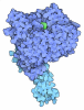
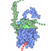
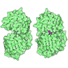

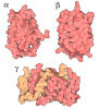
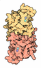



 Assembly

 Spodoptera frugiperda (fall armyworm) / References: UniProt: Q9Y297
 Mass: 24.305 Da / Num. of mol.: 1 / Source method: obtained synthetically / Formula: Mg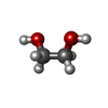
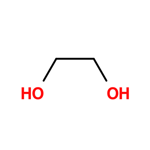 Mass: 62.068 Da / Num. of mol.: 1 / Source method: obtained synthetically / Formula: C2H6O2
 Mass: 18.015 Da / Num. of mol.: 228 / Source method: isolated from a natural source / Formula: H2O
 Sample preparation
 / Beamline: I04 / Wavelength: 0.95373 Å
 Processing