 Movie
Movie Controller


+ Open data

- Basic information
| Entry | Database: PDB / ID: 9sjj | ||||||
|---|---|---|---|---|---|---|---|
| Title | Crystal structure of SusDdex (BT3089) with bound IMO5 | ||||||
 Components Components | SusD homolog | ||||||
 Keywords Keywords | SUGAR BINDING PROTEIN / BT3089 / SusDdex / SusD / dextran / Bacteroides / glycan-binding | ||||||
| Function / homology |  Function and homology information Function and homology information | ||||||
| Biological species |  Bacteroides thetaiotaomicron VPI-5482 (bacteria) Bacteroides thetaiotaomicron VPI-5482 (bacteria) | ||||||
| Method |  X-RAY DIFFRACTION / SYNCHROTRON / MOLECULAR REPLACEMENT / Resolution: 1.7 Å X-RAY DIFFRACTION / SYNCHROTRON / MOLECULAR REPLACEMENT / Resolution: 1.7 Å | ||||||
 Authors Authors | Feasey, M. / Basle, A. / van den Berg, B. | ||||||
| Funding support |  United Kingdom, 1items United Kingdom, 1items
| ||||||
 Citation Citation | Journal: J Struct Biol X / Year: 2026 Title: Structural and functional characterisation of the dextran utilisome from . Authors: Matthew Feasey / Augustinas Silale / Arnaud Baslé / Bert van den Berg / Abstract: () is a model Bacteroidota of the healthy human gut microbiota and a specialist in glycan utilisation. Like other , has many highly regulated polysaccharide utilisation loci (PUL) that encode outer ... () is a model Bacteroidota of the healthy human gut microbiota and a specialist in glycan utilisation. Like other , has many highly regulated polysaccharide utilisation loci (PUL) that encode outer membrane (OM) TonB-dependent transporters (SusC), closely associated "lid" lipoproteins (SusD), and additional surface-exposed lipoproteins (SLPs) that bind and partially degrade specific glycans derived from host cells, diet, or other microbiota members. The canonical starch PUL products are thought to form a dynamic complex in the presence of starch. However, other PULs form stable complexes in the absence of substrate (recently named "utilisomes"), with additional surface lipoproteins tightly associated with the core SusCD complex. In this study, we characterised the dextran utilisome, with a SusCD core and an associated glycoside hydrolase (GH) and surface glycan binding protein (SBGP). Via X-ray crystallography we solved high-resolution structures of SBGP in isolation and SusD and GH bound to dextran oligosaccharides. We used isothermal titration calorimetry (ITC) to quantify ligand binding of wild type and mutant SLPs. We further used single particle cryo-EM of the catalytically inactive dextran utilisome to visualise open and closed states of the complex. Three occupied dextran binding sites were observed across SusC, SusD and GH, with substrate observed in both open and closed states of SusD. 3D variability analysis showed a minority of particles in the process of SusD lid closure. Together our work defines commonalities and differences across utilisomes dedicated to the import of simple glycans. | ||||||
| History |
|
- Structure visualization
| Structure viewer | Molecule: MolmilJmol/JSmol |
|---|
- Downloads & links
-Download
| PDBx/mmCIF format | 9sjj.cif.gz | 124.4 KB | Display | PDBx/mmCIF format |
|---|---|---|---|---|
| PDB format | pdb9sjj.ent.gz | 89 KB | Display | PDB format |
| PDBx/mmJSON format | 9sjj.json.gz | Tree view | PDBx/mmJSON format | |
| Others |  Other downloads Other downloads |
-Validation report
| Arichive directory | https://data.pdbj.org/pub/pdb/validation_reports/sj/9sjjftp://data.pdbj.org/pub/pdb/validation_reports/sj/9sjj | HTTPS FTP |
|---|
-Related structure data
| Related structure data |  9sjeC  9sjfC  9sjgC  9sjhC  9sjiC  9sjkC  9sm2C C: citing same article ( |
|---|---|
| Similar structure data |


-Links
 PDBj
PDBj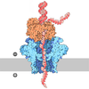
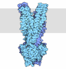
- Assembly
| Deposited unit | 
| ||||||||
|---|---|---|---|---|---|---|---|---|---|
| 1 |
| ||||||||
| Unit cell |
|
-Components
-Protein / Sugars , 2 types, 2 molecules A
| #1: Protein | Mass: 53814.082 Da / Num. of mol.: 1 Source method: isolated from a genetically manipulated source Source: (gene. exp.) Bacteroides thetaiotaomicron VPI-5482 (bacteria)Gene: BT_3089 Production host:  Escherichia coli 'BL21-Gold(DE3)pLysS AG' (bacteria) Escherichia coli 'BL21-Gold(DE3)pLysS AG' (bacteria)References: UniProt: Q8A366 |
|---|---|
| #2: Polysaccharide | alpha-D-glucopyranose-(1-6)-alpha-D-glucopyranose-(1-6)-alpha-D-glucopyranose-(1-6)-alpha-D- ...alpha-D-glucopyranose-(1-6)-alpha-D-glucopyranose-(1-6)-alpha-D-glucopyranose-(1-6)-alpha-D-glucopyranose-(1-6)-alpha-D-glucopyranose Source method: isolated from a genetically manipulated source |
-Non-polymers , 7 types, 326 molecules 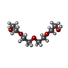
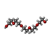
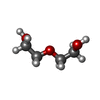

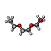

| #3: Chemical | 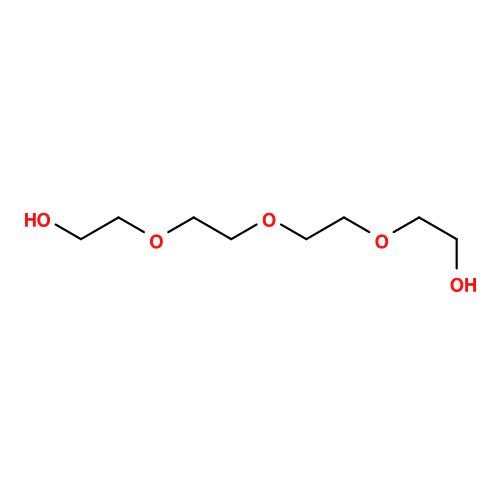 Mass: 194.226 Da / Num. of mol.: 2 / Source method: obtained synthetically / Formula: C8H18O5 / Comment: precipitant*YM Mass: 194.226 Da / Num. of mol.: 2 / Source method: obtained synthetically / Formula: C8H18O5 / Comment: precipitant*YM#4: Chemical | ChemComp-1PE / 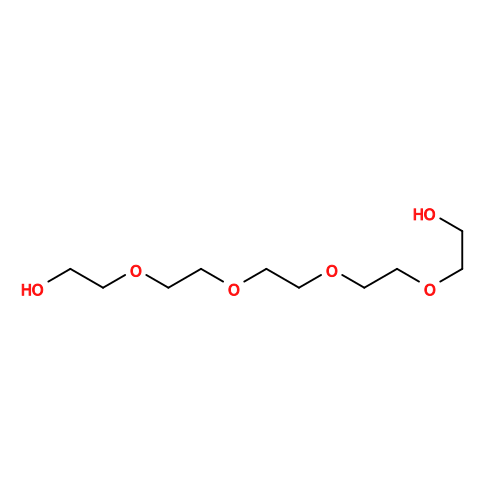 Mass: 238.278 Da / Num. of mol.: 4 / Source method: obtained synthetically / Formula: C10H22O6 / Comment: precipitant*YM Mass: 238.278 Da / Num. of mol.: 4 / Source method: obtained synthetically / Formula: C10H22O6 / Comment: precipitant*YM#5: Chemical | 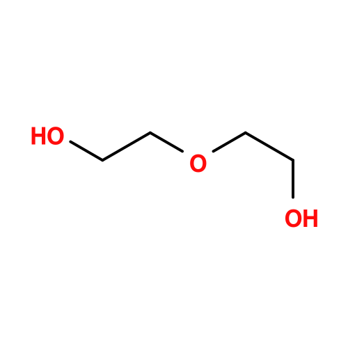 Mass: 106.120 Da / Num. of mol.: 3 / Source method: obtained synthetically / Formula: C4H10O3 Mass: 106.120 Da / Num. of mol.: 3 / Source method: obtained synthetically / Formula: C4H10O3#6: Chemical | ChemComp-P33 / |  Mass: 326.383 Da / Num. of mol.: 1 / Source method: obtained synthetically / Formula: C14H30O8 / Comment: precipitant*YM Mass: 326.383 Da / Num. of mol.: 1 / Source method: obtained synthetically / Formula: C14H30O8 / Comment: precipitant*YM#7: Chemical | ChemComp-PGE / | 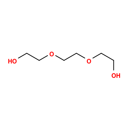 Mass: 150.173 Da / Num. of mol.: 1 / Source method: obtained synthetically / Formula: C6H14O4 Mass: 150.173 Da / Num. of mol.: 1 / Source method: obtained synthetically / Formula: C6H14O4#8: Chemical | ChemComp-SO4 / 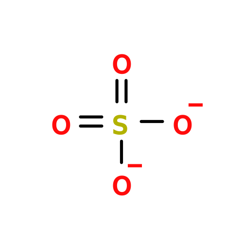 Mass: 96.063 Da / Num. of mol.: 5 / Source method: obtained synthetically / Formula: SO4 Mass: 96.063 Da / Num. of mol.: 5 / Source method: obtained synthetically / Formula: SO4#9: Water | ChemComp-HOH / | Mass: 18.015 Da / Num. of mol.: 310 / Source method: isolated from a natural source / Formula: H2O |
|---|
-Details
| Has ligand of interest | Y |
|---|---|
| Has protein modification | N |
-Experimental details
-Experiment
| Experiment | Method: X-RAY DIFFRACTION / Number of used crystals: 1 |
|---|
- Sample preparation
| Crystal | Density Matthews: 2.97 Å3/Da / Density % sol: 58.55 % |
|---|---|
| Crystal grow | Temperature: 293 K / Method: vapor diffusion, sitting drop / pH: 4.5 Details: 0.2M Lithium sulfate 0.1M Sodium acetate pH 4.5 50% w/v PEG 400 Soaked ~10 mM dextran 1.5 into grown crystals (1 crystal per drop), which cracked the large crystal. This was one fragment ...Details: 0.2M Lithium sulfate 0.1M Sodium acetate pH 4.5 50% w/v PEG 400 Soaked ~10 mM dextran 1.5 into grown crystals (1 crystal per drop), which cracked the large crystal. This was one fragment harvested for the synchrotron. |
-Data collection
| Diffraction | Mean temperature: 100 K / Serial crystal experiment: N | ||||||||||||||||||||||||||||||
|---|---|---|---|---|---|---|---|---|---|---|---|---|---|---|---|---|---|---|---|---|---|---|---|---|---|---|---|---|---|---|---|
| Diffraction source | Source: SYNCHROTRON / Site: Diamond / Beamline: I03 / Wavelength: 0.9796 Å | ||||||||||||||||||||||||||||||
| Detector | Type: DECTRIS EIGER2 XE 16M / Detector: PIXEL / Date: May 6, 2021 | ||||||||||||||||||||||||||||||
| Radiation | Protocol: SINGLE WAVELENGTH / Monochromatic (M) / Laue (L): M / Scattering type: x-ray | ||||||||||||||||||||||||||||||
| Radiation wavelength | Wavelength: 0.9796 Å / Relative weight: 1 | ||||||||||||||||||||||||||||||
| Reflection | Resolution: 1.7→67.29 Å / Num. obs: 70548 / % possible obs: 100 % / Redundancy: 25.3 % / CC1/2: 1 / Rmerge(I) obs: 0.096 / Rpim(I) all: 0.028 / Rrim(I) all: 0.1 / Χ2: 0.99 / Net I/σ(I): 19.4 | ||||||||||||||||||||||||||||||
| Reflection shell |
|
- Processing
| Software |
| |||||||||||||||||||||||||||||||||||||||||||||||||||||||||||||||||||||||||||||||||||||||||||||||||||||||||||||||||||||||||||||||||||||||||||||||||||||||||||||||||||||||||||||||||||||||||||||||||||||||||||||||||||||||||||||||||||||||
|---|---|---|---|---|---|---|---|---|---|---|---|---|---|---|---|---|---|---|---|---|---|---|---|---|---|---|---|---|---|---|---|---|---|---|---|---|---|---|---|---|---|---|---|---|---|---|---|---|---|---|---|---|---|---|---|---|---|---|---|---|---|---|---|---|---|---|---|---|---|---|---|---|---|---|---|---|---|---|---|---|---|---|---|---|---|---|---|---|---|---|---|---|---|---|---|---|---|---|---|---|---|---|---|---|---|---|---|---|---|---|---|---|---|---|---|---|---|---|---|---|---|---|---|---|---|---|---|---|---|---|---|---|---|---|---|---|---|---|---|---|---|---|---|---|---|---|---|---|---|---|---|---|---|---|---|---|---|---|---|---|---|---|---|---|---|---|---|---|---|---|---|---|---|---|---|---|---|---|---|---|---|---|---|---|---|---|---|---|---|---|---|---|---|---|---|---|---|---|---|---|---|---|---|---|---|---|---|---|---|---|---|---|---|---|---|---|---|---|---|---|---|---|---|---|---|---|---|---|---|---|---|---|
| Refinement | Method to determine structure: MOLECULAR REPLACEMENT / Resolution: 1.7→67.288 Å / Cor.coef. Fo:Fc: 0.972 / Cor.coef. Fo:Fc free: 0.954 / SU B: 1.813 / SU ML: 0.057 / Cross valid method: FREE R-VALUE / ESU R: 0.082 / ESU R Free: 0.082 / Details: Hydrogens have not been used
| |||||||||||||||||||||||||||||||||||||||||||||||||||||||||||||||||||||||||||||||||||||||||||||||||||||||||||||||||||||||||||||||||||||||||||||||||||||||||||||||||||||||||||||||||||||||||||||||||||||||||||||||||||||||||||||||||||||||
| Solvent computation | Ion probe radii: 0.8 Å / Shrinkage radii: 0.8 Å / VDW probe radii: 1.2 Å / Solvent model: MASK BULK SOLVENT | |||||||||||||||||||||||||||||||||||||||||||||||||||||||||||||||||||||||||||||||||||||||||||||||||||||||||||||||||||||||||||||||||||||||||||||||||||||||||||||||||||||||||||||||||||||||||||||||||||||||||||||||||||||||||||||||||||||||
| Displacement parameters | Biso mean: 35.229 Å2
| |||||||||||||||||||||||||||||||||||||||||||||||||||||||||||||||||||||||||||||||||||||||||||||||||||||||||||||||||||||||||||||||||||||||||||||||||||||||||||||||||||||||||||||||||||||||||||||||||||||||||||||||||||||||||||||||||||||||
| Refinement step | Cycle: LAST / Resolution: 1.7→67.288 Å
| |||||||||||||||||||||||||||||||||||||||||||||||||||||||||||||||||||||||||||||||||||||||||||||||||||||||||||||||||||||||||||||||||||||||||||||||||||||||||||||||||||||||||||||||||||||||||||||||||||||||||||||||||||||||||||||||||||||||
| Refine LS restraints |
| |||||||||||||||||||||||||||||||||||||||||||||||||||||||||||||||||||||||||||||||||||||||||||||||||||||||||||||||||||||||||||||||||||||||||||||||||||||||||||||||||||||||||||||||||||||||||||||||||||||||||||||||||||||||||||||||||||||||
| LS refinement shell | Refine-ID: X-RAY DIFFRACTION / Total num. of bins used: 20
|
