 Movie
Movie Controller

[English] 日本語
 Yorodumi
- PDB-9sdf: Structure at 1.9 A resolution of Thermus thermophilus tyrosyl-tRN... -
+ Open data

- Basic information
| Entry | Database: PDB / ID: 9sdf | |||||||||
|---|---|---|---|---|---|---|---|---|---|---|
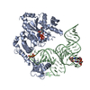
| Title | Structure at 1.9 A resolution of Thermus thermophilus tyrosyl-tRNA synthetase bound to wild-type modified tRNA(Tyr), tyrosine and AMP | |||||||||
 Components Components |
| |||||||||
 Keywords Keywords | RNA BINDING PROTEIN / aminoacyl-tRNA synthetase / ligase / tRNA | |||||||||
| Function / homology |  Function and homology information Function and homology informationtyrosyl-tRNA aminoacylation / tyrosine-tRNA ligase / tyrosine-tRNA ligase activity / RNA binding / ATP binding / cytosol Similarity search - Function | |||||||||
| Biological species |   Thermus thermophilus HB27 (bacteria) Thermus thermophilus HB27 (bacteria) | |||||||||
| Method |  X-RAY DIFFRACTION / SYNCHROTRON / MOLECULAR REPLACEMENT / Resolution: 1.9 Å X-RAY DIFFRACTION / SYNCHROTRON / MOLECULAR REPLACEMENT / Resolution: 1.9 Å | |||||||||
 Authors Authors | Cusack, S. | |||||||||
| Funding support | 1items
| |||||||||
 Citation Citation | Journal: EMBO J / Year: 2002 Title: Class I tyrosyl-tRNA synthetase has a class II mode of cognate tRNA recognition. Authors: Yaremchuk, A. / Kriklivyi, I. / Tukalo, M. / Cusack, S. | |||||||||
| History |
|
- Structure visualization
| Structure viewer | Molecule: MolmilJmol/JSmol |
|---|
- Downloads & links
-Download
| PDBx/mmCIF format | 9sdf.cif.gz | 274.8 KB | Display | PDBx/mmCIF format |
|---|---|---|---|---|
| PDB format | pdb9sdf.ent.gz | Display | PDB format | |
| PDBx/mmJSON format | 9sdf.json.gz | Tree view | PDBx/mmJSON format | |
| Others |  Other downloads Other downloads |
-Validation report
| Arichive directory | https://data.pdbj.org/pub/pdb/validation_reports/sd/9sdfftp://data.pdbj.org/pub/pdb/validation_reports/sd/9sdf | HTTPS FTP |
|---|
-Related structure data


-Links
 PDBj
PDBj


- Assembly
| Deposited unit | 
| ||||||||
|---|---|---|---|---|---|---|---|---|---|
| 1 | 
| ||||||||
| Unit cell |
|
-Components
-Protein / RNA chain , 2 types, 2 molecules AT
| #1: Protein | Mass: 48786.203 Da / Num. of mol.: 1 Source method: isolated from a genetically manipulated source Source: (gene. exp.) Thermus thermophilus HB27 (bacteria) / Gene: tyrS, TT_C1033 / Production host: |
|---|---|
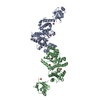
| #2: RNA chain | Mass: 27923.928 Da / Num. of mol.: 1 / Source method: isolated from a natural source / Source: (natural) Thermus thermophilus HB27 (bacteria) |
-Non-polymers , 8 types, 513 molecules 


| #3: Chemical | ChemComp-AMP /  Mass: 347.221 Da / Num. of mol.: 1 / Source method: obtained synthetically / Formula: C10H14N5O7P / Feature type: SUBJECT OF INVESTIGATION / Comment: AMP*YM Mass: 347.221 Da / Num. of mol.: 1 / Source method: obtained synthetically / Formula: C10H14N5O7P / Feature type: SUBJECT OF INVESTIGATION / Comment: AMP*YM | ||||||||||
|---|---|---|---|---|---|---|---|---|---|---|---|
| #4: Chemical | ChemComp-TYR /  Type: L-peptide linking / Mass: 181.189 Da / Num. of mol.: 1 / Source method: obtained synthetically / Formula: C9H11NO3 / Feature type: SUBJECT OF INVESTIGATION Type: L-peptide linking / Mass: 181.189 Da / Num. of mol.: 1 / Source method: obtained synthetically / Formula: C9H11NO3 / Feature type: SUBJECT OF INVESTIGATION | ||||||||||
| #5: Chemical | ChemComp-PEG /  Mass: 106.120 Da / Num. of mol.: 5 / Source method: obtained synthetically / Formula: C4H10O3 Mass: 106.120 Da / Num. of mol.: 5 / Source method: obtained synthetically / Formula: C4H10O3#6: Chemical |  Mass: 96.063 Da / Num. of mol.: 2 / Source method: obtained synthetically / Formula: SO4 Mass: 96.063 Da / Num. of mol.: 2 / Source method: obtained synthetically / Formula: SO4#7: Chemical | ChemComp-1PE /  Mass: 238.278 Da / Num. of mol.: 5 / Source method: obtained synthetically / Formula: C10H22O6 / Comment: precipitant*YM Mass: 238.278 Da / Num. of mol.: 5 / Source method: obtained synthetically / Formula: C10H22O6 / Comment: precipitant*YM#8: Chemical | ChemComp-PGE / |  Mass: 150.173 Da / Num. of mol.: 1 / Source method: obtained synthetically / Formula: C6H14O4 Mass: 150.173 Da / Num. of mol.: 1 / Source method: obtained synthetically / Formula: C6H14O4#9: Chemical | ChemComp-MG / |  Mass: 24.305 Da / Num. of mol.: 1 / Source method: obtained synthetically / Formula: Mg Mass: 24.305 Da / Num. of mol.: 1 / Source method: obtained synthetically / Formula: Mg#10: Water | ChemComp-HOH / | Mass: 18.015 Da / Num. of mol.: 497 / Source method: isolated from a natural source / Formula: H2O |
-Details
| Has ligand of interest | Y |
|---|---|
| Has protein modification | N |
-Experimental details
-Experiment
| Experiment | Method: X-RAY DIFFRACTION / Number of used crystals: 1 |
|---|
- Sample preparation
| Crystal | Density Matthews: 2.89 Å3/Da / Density % sol: 57.48 % |
|---|---|
| Crystal grow | Temperature: 293 K / Method: vapor diffusion, hanging drop / Details: do not know Cryo: 22% PEG 400 |
-Data collection
| Diffraction | Mean temperature: 100 K / Serial crystal experiment: N |
|---|---|
| Diffraction source | Source: SYNCHROTRON / Site: ESRF  / Beamline: BM14 / Wavelength: 0.97625 Å / Beamline: BM14 / Wavelength: 0.97625 Å |
| Detector | Type: MAR CCD 300 mm / Detector: CCD / Date: Aug 29, 2002 |
| Radiation | Protocol: SINGLE WAVELENGTH / Monochromatic (M) / Laue (L): M / Scattering type: x-ray |
| Radiation wavelength | Wavelength: 0.97625 Å / Relative weight: 1 |
| Reflection | Resolution: 1.9→22.44 Å / Num. obs: 127578 / % possible obs: 99.1 % / Redundancy: 2.48 % / Rmerge(I) obs: 0.099 / Net I/σ(I): 8.18 |
| Reflection shell | Resolution: 1.9→1.95 Å / Rmerge(I) obs: 0.518 / Num. unique obs: 5005 / % possible all: 99.1 |
- Processing
| Software |
| |||||||||||||||||||||||||||||||||||||||||||||||||||||||||||||||||||||||||||||||||||||||||||||||||||||||||||||||||||||||||||||||||||||||||||||||||||||||||||||||||||||||||||||||||||||||||||||||||||||||||||||||||||||||||
|---|---|---|---|---|---|---|---|---|---|---|---|---|---|---|---|---|---|---|---|---|---|---|---|---|---|---|---|---|---|---|---|---|---|---|---|---|---|---|---|---|---|---|---|---|---|---|---|---|---|---|---|---|---|---|---|---|---|---|---|---|---|---|---|---|---|---|---|---|---|---|---|---|---|---|---|---|---|---|---|---|---|---|---|---|---|---|---|---|---|---|---|---|---|---|---|---|---|---|---|---|---|---|---|---|---|---|---|---|---|---|---|---|---|---|---|---|---|---|---|---|---|---|---|---|---|---|---|---|---|---|---|---|---|---|---|---|---|---|---|---|---|---|---|---|---|---|---|---|---|---|---|---|---|---|---|---|---|---|---|---|---|---|---|---|---|---|---|---|---|---|---|---|---|---|---|---|---|---|---|---|---|---|---|---|---|---|---|---|---|---|---|---|---|---|---|---|---|---|---|---|---|---|---|---|---|---|---|---|---|---|---|---|---|---|---|---|---|---|
| Refinement | Method to determine structure: MOLECULAR REPLACEMENT / Resolution: 1.9→16.74 Å / SU ML: 0.19 / Cross valid method: FREE R-VALUE / σ(F): 1.05 / Phase error: 20.89 / Stereochemistry target values: ML
| |||||||||||||||||||||||||||||||||||||||||||||||||||||||||||||||||||||||||||||||||||||||||||||||||||||||||||||||||||||||||||||||||||||||||||||||||||||||||||||||||||||||||||||||||||||||||||||||||||||||||||||||||||||||||
| Solvent computation | Shrinkage radii: 0.9 Å / VDW probe radii: 1.1 Å / Solvent model: FLAT BULK SOLVENT MODEL | |||||||||||||||||||||||||||||||||||||||||||||||||||||||||||||||||||||||||||||||||||||||||||||||||||||||||||||||||||||||||||||||||||||||||||||||||||||||||||||||||||||||||||||||||||||||||||||||||||||||||||||||||||||||||
| Refinement step | Cycle: LAST / Resolution: 1.9→16.74 Å
| |||||||||||||||||||||||||||||||||||||||||||||||||||||||||||||||||||||||||||||||||||||||||||||||||||||||||||||||||||||||||||||||||||||||||||||||||||||||||||||||||||||||||||||||||||||||||||||||||||||||||||||||||||||||||
| Refine LS restraints |
| |||||||||||||||||||||||||||||||||||||||||||||||||||||||||||||||||||||||||||||||||||||||||||||||||||||||||||||||||||||||||||||||||||||||||||||||||||||||||||||||||||||||||||||||||||||||||||||||||||||||||||||||||||||||||
| LS refinement shell |
|
