[English] 日本語
Yorodumi
- PDB-9s6j: HIV-1 capsid (M-group) - CPSF6 -

+
Open data


ID or keywords:

Loading...

-
Basic information

Entry
Database: PDB / ID: 9s6j
TitleHIV-1 capsid (M-group) - CPSF6
Components
  • Cleavage and polyadenylation specificity factor subunit 6
  • Gag polyprotein
KeywordsVIRAL PROTEIN / Hexameric HIV-1 (M-group)
Function / homology
Function and homology information


exon-exon junction complex binding / mRNA alternative polyadenylation / positive regulation of RNA export from nucleus / mRNA cleavage factor complex / interchromatin granule / co-transcriptional mRNA 3'-end processing, cleavage and polyadenylation pathway / perichromatin fibrils / Processing of Intronless Pre-mRNAs / mRNA cleavage and polyadenylation specificity factor complex / mRNA 3'-end processing ...exon-exon junction complex binding / mRNA alternative polyadenylation / positive regulation of RNA export from nucleus / mRNA cleavage factor complex / interchromatin granule / co-transcriptional mRNA 3'-end processing, cleavage and polyadenylation pathway / perichromatin fibrils / Processing of Intronless Pre-mRNAs / mRNA cleavage and polyadenylation specificity factor complex / mRNA 3'-end processing / Signaling by cytosolic FGFR1 fusion mutants / paraspeckles / mRNA 3'-end processing / RNA Polymerase II Transcription Termination / protein heterotetramerization / ribosomal large subunit binding / Signaling by FGFR1 in disease / protein tetramerization / host multivesicular body / mRNA processing / mRNA Polyadenylation / viral nucleocapsid / Dengue Virus-Host Interactions / nuclear speck / ribonucleoprotein complex / viral translational frameshifting / mRNA binding / host cell nucleus / host cell plasma membrane / virion membrane / structural molecule activity / RNA binding / nucleoplasm / zinc ion binding / ATP binding / membrane / nucleus / cytoplasm
Similarity search - Function
Cleavage and polyadenylation specificity factor subunit 6 / CPSF6/7 family / : / CPSF6-like, RSLD domain, N-terminal region / : / gag protein p24 N-terminal domain / RNA recognition motif / RNA recognition motif / Eukaryotic RNA Recognition Motif (RRM) profile. / RNA recognition motif domain ...Cleavage and polyadenylation specificity factor subunit 6 / CPSF6/7 family / : / CPSF6-like, RSLD domain, N-terminal region / : / gag protein p24 N-terminal domain / RNA recognition motif / RNA recognition motif / Eukaryotic RNA Recognition Motif (RRM) profile. / RNA recognition motif domain / RNA-binding domain superfamily / Immunodeficiency lentiviral matrix, N-terminal / gag gene protein p17 (matrix protein) / Matrix protein, lentiviral and alpha-retroviral, N-terminal / Retroviral nucleocapsid Gag protein p24, C-terminal domain / Gag protein p24 C-terminal domain / Retrovirus capsid, C-terminal / Retroviral matrix protein / Retrovirus capsid, N-terminal / Zinc knuckle / Zinc finger, CCHC-type superfamily / Zinc finger, CCHC-type / Zinc finger CCHC-type profile. / Nucleotide-binding alpha-beta plait domain superfamily
Similarity search - Domain/homology
IODIDE ION / Gag polyprotein / Cleavage and polyadenylation specificity factor subunit 6
Similarity search - Component
Biological speciesHuman immunodeficiency virus type 1
Homo sapiens (human)
MethodX-RAY DIFFRACTION / SYNCHROTRON / MOLECULAR REPLACEMENT / Resolution: 2.4 Å
AuthorsGovasli, M.A.L. / Pinotsis, N. / Towers, G. / Selwood, D. / Jacques, D.A.
Funding support United Kingdom, European Union, Australia, 4items
OrganizationGrant numberCountry
Wellcome Trust220863 United Kingdom
Wellcome Trust214344 United Kingdom
European Research Council (ERC)339223European Union
National Health and Medical Research Council (NHMRC, Australia)GNT2013215 Australia
CitationJournal: To Be Published
Title: Cofactor-mimicking HIV-1 capsid inhibitors, and their escape mutants, drive innate immune sensing
Authors: Govasli, M.A.L. / Pinotsis, N. / Towers, G. / Selwood, D. / Jacques, D.A.
History
DepositionAug 1, 2025Deposition site: PDBE / Processing site: PDBE
Revision 1.0Aug 12, 2026Provider: repository / Type: Initial release

-
Structure visualization

Structure viewerMolecule:
MolmilJmol/JSmol

Downloads & links

-
Assembly

Deposited unit
A: Gag polyprotein
B: Cleavage and polyadenylation specificity factor subunit 6
hetero molecules


Theoretical massNumber of molelcules
Total (without water)27,43710
Polymers26,9142
Non-polymers5238
Water61334
1


  • Idetical with deposited unit
  • defined by author
  • Evidence: gel filtration
TypeNameSymmetry operationNumber
identity operation1_555x,y,z1
Buried area2170 Å2
ΔGint-55 kcal/mol
Surface area13970 Å2
Unit cell
Length a, b, c (Å)92.67, 92.67, 57.98
Angle α, β, γ (deg.)90, 90, 120
Int Tables number168
Space group name H-MP6

-
Components

-
Protein / Protein/peptide , 2 types, 2 molecules AB

#1: Protein Gag polyprotein


Mass: 25630.426 Da / Num. of mol.: 1
Source method: isolated from a genetically manipulated source
Source: (gene. exp.) Human immunodeficiency virus type 1 (NEW YORK-5 ISOLATE)
Gene: gag / Production host: Escherichia coli BL21(DE3) (bacteria) / References: UniProt: B6DRA0
#2: Protein/peptide Cleavage and polyadenylation specificity factor subunit 6 / Cleavage and polyadenylation specificity factor 68 kDa subunit / CPSF 68 kDa subunit / Cleavage ...Cleavage and polyadenylation specificity factor 68 kDa subunit / CPSF 68 kDa subunit / Cleavage factor Im complex 68 kDa subunit / CFIm68 / Pre-mRNA cleavage factor Im 68 kDa subunit / Protein HPBRII-4/7


Mass: 1283.472 Da / Num. of mol.: 1 / Source method: obtained synthetically / Source: (synth.) Homo sapiens (human) / References: UniProt: Q16630

-
Non-polymers , 4 types, 42 molecules

#3: Chemical ChemComp-IOD / IODIDE ION


Mass: 126.904 Da / Num. of mol.: 2 / Source method: obtained synthetically / Formula: I
#4: Chemical
ChemComp-CL / CHLORIDE ION


Mass: 35.453 Da / Num. of mol.: 5 / Source method: obtained synthetically / Formula: Cl
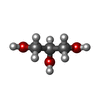
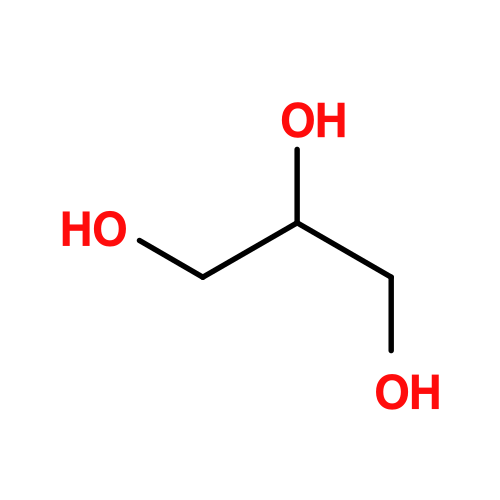
#5: Chemical ChemComp-GOL / GLYCEROL / GLYCERIN / PROPANE-1,2,3-TRIOL


Mass: 92.094 Da / Num. of mol.: 1 / Source method: obtained synthetically / Formula: C3H8O3
#6: Water ChemComp-HOH / water


Mass: 18.015 Da / Num. of mol.: 34 / Source method: isolated from a natural source / Formula: H2O

-
Details

Has ligand of interestN
Has protein modificationY

-
Experimental details

-
Experiment

ExperimentMethod: X-RAY DIFFRACTION / Number of used crystals: 1

-
Sample preparation

CrystalDensity Matthews: 2.67 Å3/Da / Density % sol: 53.94 %
Crystal growTemperature: 298 K / Method: vapor diffusion, hanging drop / pH: 6.5
Details: 9.5-11% PEG 3350 (v/v), 250-350 mM NaI, 100 mM Sodium Cacodylate [pH 6.5]. Crystals grew in 1 uL protein (3 mg/mL) + 1 uL crystallant. Cryoprotected in 20% (v/v) Glycerol.

-
Data collection

DiffractionMean temperature: 100 K / Serial crystal experiment: N
Diffraction sourceSource: SYNCHROTRON / Site: EMBL/DESY, HAMBURG / Beamline: X13 / Wavelength: 0.97626 Å
DetectorType: DECTRIS EIGER X 16M / Detector: PIXEL / Date: Aug 10, 2019
RadiationMonochromator: M / Protocol: SINGLE WAVELENGTH / Monochromatic (M) / Laue (L): M / Scattering type: x-ray
Radiation wavelengthWavelength: 0.97626 Å / Relative weight: 1
ReflectionResolution: 2.4→80.26 Å / Num. obs: 11179 / % possible obs: 99.4 % / Redundancy: 4.5 % / CC1/2: 0.998 / Rmerge(I) obs: 0.094 / Net I/σ(I): 9.33
Reflection shellResolution: 2.4→2.46 Å / Num. unique obs: 816 / CC1/2: 0.315

-
Processing

Software
NameVersionClassification
BUSTER2.10.3refinement
XDS1.0.5 (20200319)data reduction
XSCALEMar 15, 2019 BUILT=20190315data scaling
PHASER2.8.3phasing
RefinementMethod to determine structure: MOLECULAR REPLACEMENT / Resolution: 2.4→80.25 Å / Cor.coef. Fo:Fc: 0.933 / Cor.coef. Fo:Fc free: 0.902 / SU R Cruickshank DPI: 0.399 / Cross valid method: THROUGHOUT / SU R Blow DPI: 0.384 / SU Rfree Blow DPI: 0.258 / SU Rfree Cruickshank DPI: 0.264
RfactorNum. reflection% reflectionSelection details
Rfree0.2691 559 -RANDOM
Rwork0.2331 ---
obs0.2349 11179 99.4 %-
Displacement parametersBiso mean: 87.52 Å2
Baniso -1Baniso -2Baniso -3
1-6.8532 Å20 Å20 Å2
2--6.8532 Å20 Å2
3----13.7064 Å2
Refine analyzeLuzzati coordinate error obs: 0.45 Å
Refinement stepCycle: LAST / Resolution: 2.4→80.25 Å
ProteinNucleic acidLigandSolventTotal
Num. atoms1812 0 13 34 1859
Refine LS restraints
Refine-IDTypeDev idealNumberRestraint functionWeight
X-RAY DIFFRACTIONt_bond_d0.0051882HARMONIC2
X-RAY DIFFRACTIONt_angle_deg0.722562HARMONIC2
X-RAY DIFFRACTIONt_dihedral_angle_d647SINUSOIDAL2
X-RAY DIFFRACTIONt_gen_planes317HARMONIC5
X-RAY DIFFRACTIONt_it1882HARMONIC10
X-RAY DIFFRACTIONt_chiral_improper_torsion249SEMIHARMONIC5
X-RAY DIFFRACTIONt_sum_occupancies2HARMONIC1
X-RAY DIFFRACTIONt_ideal_dist_contact1406SEMIHARMONIC4
X-RAY DIFFRACTIONt_omega_torsion1.89
X-RAY DIFFRACTIONt_other_torsion17.04
LS refinement shellResolution: 2.4→2.43 Å
RfactorNum. reflection% reflection
Rfree0.3089 20 -
Rwork0.2422 --
obs0.2455 415 100 %
Refinement TLS params.

Refine-ID: X-RAY DIFFRACTION

IDL112)L122)L132)L222)L232)L332)S11 (Å °)S12 (Å °)S13 (Å °)S21 (Å °)S22 (Å °)S23 (Å °)S31 (Å °)S32 (Å °)S33 (Å °)T112)T122)T132)T222)T232)T332)Origin x (Å)Origin y (Å)Origin z (Å)
11.91560.8527-0.2422.1704-0.65290-0.1582-0.16350.1191-0.1635-0.14620.10180.11910.10180.3045-0.13250.06720.0031-0.1874-0.06260.2359-0.3193-16.8452-3.4598
20.89990.93460.11351.67370.91573.6704-0.1368-0.41280.347-0.41280.259-0.12180.347-0.1218-0.12220.0103-0.00760.1991-0.25-0.06060.11152.2927-31.2503-12.7793
31.24550.3537-0.82173.70410.02412.12930.3220.32310.66620.3231-0.3624-0.21830.6662-0.21830.0404-0.0092-0.29680.0631-0.0175-0.1266-0.1128-26.8407-28.249213.3833
44.229-4.4144-0.50912.60513.066700.1024-0.59450.1085-0.59450.00090.69110.10850.6911-0.10340.08450.13520.12790.04980.0049-0.202911.6285-32.1974-2.7672
Refinement TLS group
IDRefine-IDRefine TLS-IDSelection detailsAuth asym-IDAuth seq-ID
1X-RAY DIFFRACTION1{ A|1 - A|62}A1 - 62
2X-RAY DIFFRACTION2{ A|63 - A|142}A63 - 142
3X-RAY DIFFRACTION3{ A|143 - A|221}A143 - 221
4X-RAY DIFFRACTION4{ B|313 - B|324}B313 - 324

+
About Yorodumi

-
News

-
Feb 9, 2022. New format data for meta-information of EMDB entries

New format data for meta-information of EMDB entries

  • Version 3 of the EMDB header file is now the official format.
  • The previous official version 1.9 will be removed from the archive.

Related info.:EMDB header

External links:wwPDB to switch to version 3 of the EMDB data model

-
Aug 12, 2020. Covid-19 info

Covid-19 info

URL: https://pdbj.org/emnavi/covid19.php

New page: Covid-19 featured information page in EM Navigator.

Related info.:Covid-19 info / Mar 5, 2020. Novel coronavirus structure data

+
Mar 5, 2020. Novel coronavirus structure data

Novel coronavirus structure data

Related info.:Yorodumi Speices / Aug 12, 2020. Covid-19 info

External links:COVID-19 featured content - PDBj / Molecule of the Month (242):Coronavirus Proteases

+
Jan 31, 2019. EMDB accession codes are about to change! (news from PDBe EMDB page)

EMDB accession codes are about to change! (news from PDBe EMDB page)

  • The allocation of 4 digits for EMDB accession codes will soon come to an end. Whilst these codes will remain in use, new EMDB accession codes will include an additional digit and will expand incrementally as the available range of codes is exhausted. The current 4-digit format prefixed with “EMD-” (i.e. EMD-XXXX) will advance to a 5-digit format (i.e. EMD-XXXXX), and so on. It is currently estimated that the 4-digit codes will be depleted around Spring 2019, at which point the 5-digit format will come into force.
  • The EM Navigator/Yorodumi systems omit the EMD- prefix.

Related info.:Q: What is EMD? / ID/Accession-code notation in Yorodumi/EM Navigator

External links:EMDB Accession Codes are Changing Soon! / Contact to PDBj

+
Jul 12, 2017. Major update of PDB

Major update of PDB

  • wwPDB released updated PDB data conforming to the new PDBx/mmCIF dictionary.
  • This is a major update changing the version number from 4 to 5, and with Remediation, in which all the entries are updated.
  • In this update, many items about electron microscopy experimental information are reorganized (e.g. em_software).
  • Now, EM Navigator and Yorodumi are based on the updated data.

External links:wwPDB Remediation / Enriched Model Files Conforming to OneDep Data Standards Now Available in the PDB FTP Archive

-
Yorodumi

Thousand views of thousand structures

  • Yorodumi is a browser for structure data from EMDB, PDB, SASBDB, etc.
  • This page is also the successor to EM Navigator detail page, and also detail information page/front-end page for Omokage search.
  • The word "yorodu" (or yorozu) is an old Japanese word meaning "ten thousand". "mi" (miru) is to see.

Related info.:EMDB / PDB / SASBDB / Comparison of 3 databanks / Yorodumi Search / Aug 31, 2016. New EM Navigator & Yorodumi / Yorodumi Papers / Jmol/JSmol / Function and homology information / Changes in new EM Navigator and Yorodumi

Read more