 Movie
Movie Controller


+ Open data

- Basic information
| Entry | Database: PDB / ID: 9rx8 | ||||||||||||||||||||||||
|---|---|---|---|---|---|---|---|---|---|---|---|---|---|---|---|---|---|---|---|---|---|---|---|---|---|
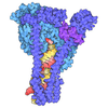
| Title | Apo VPS34-CII (VPS34/VPS15/BECLIN1/UVRAG) | ||||||||||||||||||||||||
 Components Components |
| ||||||||||||||||||||||||
 Keywords Keywords | ENDOCYTOSIS / Lipid kinase / GTPase / kinase / autophagy / endocytic trafficking / SIGNALING PROTEIN | ||||||||||||||||||||||||
| Function / homology |  Function and homology information Function and homology informationprotein-containing complex organization / nucleus-vacuole junction / cellular response to aluminum ion / positive regulation of protein lipidation / positive regulation of stress granule assembly / postsynaptic endosome / Toll Like Receptor 9 (TLR9) Cascade / Synthesis of PIPs at the late endosome membrane / phosphatidylinositol 3-kinase complex, class III / cellular response to oxygen-glucose deprivation ...protein-containing complex organization / nucleus-vacuole junction / cellular response to aluminum ion / positive regulation of protein lipidation / positive regulation of stress granule assembly / postsynaptic endosome / Toll Like Receptor 9 (TLR9) Cascade / Synthesis of PIPs at the late endosome membrane / phosphatidylinositol 3-kinase complex, class III / cellular response to oxygen-glucose deprivation / Synthesis of PIPs at the early endosome membrane / phosphatidylinositol 3-kinase complex, class III, type II / phosphatidylinositol 3-kinase complex, class III, type I / response to mitochondrial depolarisation / presynaptic endosome / positive regulation of attachment of mitotic spindle microtubules to kinetochore / host-mediated activation of viral genome replication / engulfment of apoptotic cell / negative regulation of lysosome organization / phosphatidylinositol kinase activity / SMAD protein signal transduction / Synthesis of PIPs at the Golgi membrane / early endosome to late endosome transport / receptor catabolic process / response to L-leucine / cytoplasmic side of mitochondrial outer membrane / positive regulation of autophagosome assembly / protein targeting to lysosome / protein targeting to vacuole / late endosome to vacuole transport / endosome organization / pexophagy / Dengue virus modulates apoptosis / positive regulation of natural killer cell mediated cytotoxicity / Translation of Replicase and Assembly of the Replication Transcription Complex / phagophore assembly site / cellular response to nitrogen starvation / centrosome cycle / negative regulation of programmed cell death / phosphatidylinositol 3-kinase / phosphatidylinositol-3-phosphate biosynthetic process / 1-phosphatidylinositol-3-kinase activity / response to vitamin E / Macroautophagy / p38MAPK cascade / response to iron(II) ion / RSV-host interactions / cytoplasmic pattern recognition receptor signaling pathway / phosphatidylinositol phosphate biosynthetic process / phosphatidylinositol-mediated signaling / autolysosome / PI3K Cascade / autophagosome maturation / RHO GTPases Activate NADPH Oxidases / chromosome, centromeric region / JNK cascade / mitotic metaphase chromosome alignment / autophagosome assembly / axoneme / mitophagy / cellular response to glucose starvation / synaptic vesicle endocytosis / cellular defense response / phosphatidylinositol 3-kinase binding / regulation of macroautophagy / positive regulation of intrinsic apoptotic signaling pathway / vesicle-mediated transport / phagocytic vesicle / positive regulation of autophagy / cellular response to epidermal growth factor stimulus / autophagosome / cellular response to copper ion / cellular response to amino acid starvation / macroautophagy / phosphatidylinositol 3-kinase/protein kinase B signal transduction / regulation of cytokinesis / protein processing / Antigen Presentation: Folding, assembly and peptide loading of class I MHC / circadian rhythm / trans-Golgi network / regulation of autophagy / response to lead ion / endocytosis / cellular response to hydrogen peroxide / ISG15 antiviral mechanism / GABA-ergic synapse / phagocytic vesicle membrane / autophagy / late endosome / peroxisome / protein-containing complex assembly / GTPase binding / midbody / Translation of Replicase and Assembly of the Replication Transcription Complex / defense response to virus / positive regulation of phosphatidylinositol 3-kinase/protein kinase B signal transduction / response to hypoxia / molecular adaptor activity / early endosome / nuclear body Similarity search - Function | ||||||||||||||||||||||||
| Biological species |  Homo sapiens (human) Homo sapiens (human) | ||||||||||||||||||||||||
| Method | ELECTRON MICROSCOPY / single particle reconstruction / cryo EM / Resolution: 3.87 Å | ||||||||||||||||||||||||
 Authors Authors | Spokaite, S. / Ohashi, Y. / Dessus, A.N. / Bourguet, M. / Williams, R.L. | ||||||||||||||||||||||||
| Funding support |  United Kingdom, 2items United Kingdom, 2items
| ||||||||||||||||||||||||
 Citation Citation | Journal: Elife / Year: 2026 Title: A novel RAB5 binding site in human VPS34-CII that is likely the primordial site in eukaryotic evolution. Authors: Saule Spokaite / Yohei Ohashi / Maxime Bourguet / Antoine Nicolas Dessus / Roger L Williams / Abstract: RAB5-GTP activation of the multiprotein VPS34 complex II (VPS34-CII) is critical for endosomal sorting and maturation, phagocytosis, and receptor downregulation. RAB5-GTP activates VPS34-CII by ...RAB5-GTP activation of the multiprotein VPS34 complex II (VPS34-CII) is critical for endosomal sorting and maturation, phagocytosis, and receptor downregulation. RAB5-GTP activates VPS34-CII by binding to a helical insertion in the C2 domain of VPS34 on the BECLIN1/UVRAG-containing adaptor arm of the complex. The autophagy complex, VPS34 complex I (VPS34-CI), features a unique ATG14L subunit in place of the VPS34-CII UVRAG subunit, and we found that this distorts the adaptor arm to alter the VPS34 RAB-GTPase binding pocket so that it preferentially binds RAB1-GTP. Surprisingly, our higher-resolution single-particle cryo-EM structure of VPS34-CII showed a second RAB5-GTP binding site on the VPS15 solenoid region. This site (VPS15-RAB5-site) appears to be the primordial RAB5-binding region. A mutant in the helical insertion of the C2 domain of human VPS34 that mimics the sequence abolishes RAB5 binding to VPS34. Mutation of the VPS15-RAB5-site ortholog in VPS15 resulted in defective CPY sorting, loss of colocalisation with the RAB5 ortholog Vps21, and loss of binding to Vps21 in vitro. Evolutionary expansion from one to two RAB5-orthologue binding sites may have increased membrane binding and VPS34-CII activity to adapt to more complex endocytic systems. #1: Journal: Elife / Year: 2026Title: A novel RAB5 binding site in human VPS34-CII that is likely the primordial site in eukaryotic evolution Authors: Spokaite, S. / Ohashi, Y. / Bourguet, M. / Dessus, A.N. / Williams, R.L. | ||||||||||||||||||||||||
| History |
|
- Structure visualization
| Structure viewer | Molecule: MolmilJmol/JSmol |
|---|
- Downloads & links
-Download
| PDBx/mmCIF format | 9rx8.cif.gz | 526.2 KB | Display | PDBx/mmCIF format |
|---|---|---|---|---|
| PDB format | pdb9rx8.ent.gz | 415.1 KB | Display | PDB format |
| PDBx/mmJSON format | 9rx8.json.gz | Tree view | PDBx/mmJSON format | |
| Others |  Other downloads Other downloads |
-Validation report
| Arichive directory | https://data.pdbj.org/pub/pdb/validation_reports/rx/9rx8ftp://data.pdbj.org/pub/pdb/validation_reports/rx/9rx8 | HTTPS FTP |
|---|
-Related structure data
| Related structure data |  54361MC  9rx5C  9rx6C  9rx9C  9rxaC  9rxbC  9s47C M: map data used to model this data C: citing same article ( |
|---|---|
| Similar structure data |


-Links
 PDBj
PDBj





- Assembly
| Deposited unit | 
|
|---|---|
| 1 |
|
-Components
-Protein , 4 types, 4 molecules ABCD
| #1: Protein | Mass: 101680.328 Da / Num. of mol.: 1 Source method: isolated from a genetically manipulated source Source: (gene. exp.) Homo sapiens (human) / Gene: PIK3C3, VPS34 / Cell line (production host): Expi293 / Production host: Homo sapiens (human) / References: UniProt: Q8NEB9, phosphatidylinositol 3-kinase |
|---|---|
| #2: Protein | Mass: 154659.188 Da / Num. of mol.: 1 Source method: isolated from a genetically manipulated source Details: Full length VPS15 with 13 extra residues left after TEV cleavage (VPS15-SRPTTASENLYFQ) Source: (gene. exp.) Homo sapiens (human) / Gene: PIK3R4, VPS15 / Cell line (production host): Expi293F / Production host: Homo sapiens (human)References: UniProt: Q99570, non-specific serine/threonine protein kinase |
| #3: Protein | Mass: 51953.102 Da / Num. of mol.: 1 Source method: isolated from a genetically manipulated source Source: (gene. exp.) Homo sapiens (human) / Gene: BECN1, GT197 / Production host: Homo sapiens (human) / References: UniProt: Q14457 |
| #4: Protein | Mass: 78289.852 Da / Num. of mol.: 1 / Mutation: P10Q Source method: isolated from a genetically manipulated source Details: Full length UVRAG with a P10Q mutation / Source: (gene. exp.) Homo sapiens (human) / Gene: UVRAG / Production host: Homo sapiens (human) / References: UniProt: Q6P1X0 |
-Non-polymers , 4 types, 4 molecules 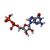


| #5: Chemical | ChemComp-GDP / 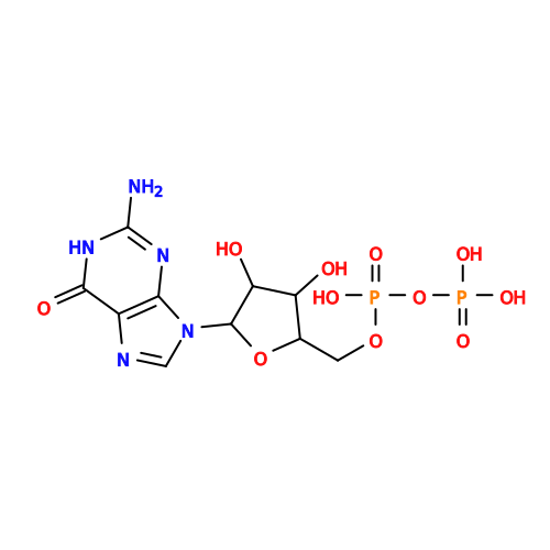 Type: RNA linking / Mass: 443.201 Da / Num. of mol.: 1 / Source method: obtained synthetically / Formula: C10H15N5O11P2 / Feature type: SUBJECT OF INVESTIGATION / Comment: GDP, energy-carrying molecule*YM Type: RNA linking / Mass: 443.201 Da / Num. of mol.: 1 / Source method: obtained synthetically / Formula: C10H15N5O11P2 / Feature type: SUBJECT OF INVESTIGATION / Comment: GDP, energy-carrying molecule*YM |
|---|---|
| #6: Chemical | ChemComp-MG /  Mass: 24.305 Da / Num. of mol.: 1 / Source method: obtained synthetically / Formula: Mg / Feature type: SUBJECT OF INVESTIGATION Mass: 24.305 Da / Num. of mol.: 1 / Source method: obtained synthetically / Formula: Mg / Feature type: SUBJECT OF INVESTIGATION |
| #7: Chemical | ChemComp-MYR / 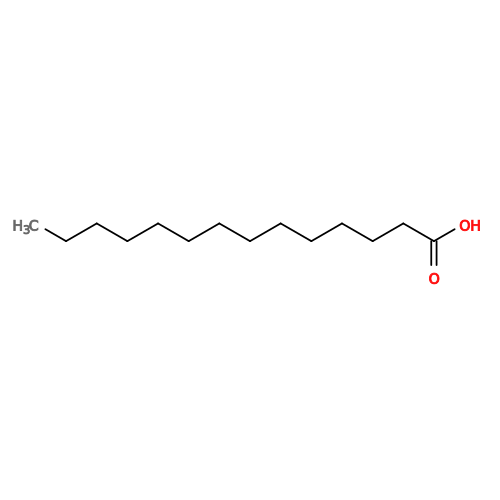 Mass: 228.371 Da / Num. of mol.: 1 / Source method: obtained synthetically / Formula: C14H28O2 / Feature type: SUBJECT OF INVESTIGATION Mass: 228.371 Da / Num. of mol.: 1 / Source method: obtained synthetically / Formula: C14H28O2 / Feature type: SUBJECT OF INVESTIGATION |
| #8: Chemical | ChemComp-ZN /  Mass: 65.409 Da / Num. of mol.: 1 / Source method: obtained synthetically / Formula: Zn / Feature type: SUBJECT OF INVESTIGATION Mass: 65.409 Da / Num. of mol.: 1 / Source method: obtained synthetically / Formula: Zn / Feature type: SUBJECT OF INVESTIGATION |
-Details
| Has ligand of interest | Y |
|---|---|
| Has protein modification | Y |
-Experimental details
-Experiment
| Experiment | Method: ELECTRON MICROSCOPY |
|---|---|
| EM experiment | Aggregation state: PARTICLE / 3D reconstruction method: single particle reconstruction |
- Sample preparation
| Component | Name: Human VPS34-CII (VPS34/VPS15/BECLIN1/UVRAG) incubated with 22-fold molar excess of RAB5A-GTP (C19S, C63S, Q79L) Type: COMPLEX Details: No RAB5A molecules were bound to VPS34-CII in this class Entity ID: #1, #4, #3 / Source: RECOMBINANT | ||||||||||||||||||||||||||||||||||||||||
|---|---|---|---|---|---|---|---|---|---|---|---|---|---|---|---|---|---|---|---|---|---|---|---|---|---|---|---|---|---|---|---|---|---|---|---|---|---|---|---|---|---|
| Molecular weight | Value: 0.432 MDa / Experimental value: NO | ||||||||||||||||||||||||||||||||||||||||
| Source (natural) | Organism: Homo sapiens (human) | ||||||||||||||||||||||||||||||||||||||||
| Source (recombinant) | Organism: Homo sapiens (human) | ||||||||||||||||||||||||||||||||||||||||
| Buffer solution | pH: 8 | ||||||||||||||||||||||||||||||||||||||||
| Buffer component |
| ||||||||||||||||||||||||||||||||||||||||
| Specimen | Conc.: 1.5 mg/ml / Embedding applied: NO / Shadowing applied: NO / Staining applied: NO / Vitrification applied: YES Details: Sample was crosslinked with 20 uM BS3 for 20 minutes on ice, then quenched with 100 mM TRIS pH 8.0 prior to vitrification | ||||||||||||||||||||||||||||||||||||||||
| Specimen support | Grid material: GOLD / Grid mesh size: 300 divisions/in. / Grid type: UltrAuFoil R1.2/1.3 | ||||||||||||||||||||||||||||||||||||||||
| Vitrification | Instrument: FEI VITROBOT MARK III / Cryogen name: ETHANE / Humidity: 100 % / Chamber temperature: 287 K Details: 3.5 uL sample, 0 second wait, 4 second blot, blot force +8 |
- Electron microscopy imaging
| Experimental equipment |  Model: Titan Krios / Image courtesy: FEI Company |
|---|---|
| Microscopy | Model: TFS KRIOS |
| Electron gun | Electron source:  FIELD EMISSION GUN / Accelerating voltage: 300 kV / Illumination mode: FLOOD BEAM FIELD EMISSION GUN / Accelerating voltage: 300 kV / Illumination mode: FLOOD BEAM |
| Electron lens | Mode: BRIGHT FIELD / Nominal magnification: 105000 X / Nominal defocus max: 2200 nm / Nominal defocus min: 800 nm / C2 aperture diameter: 50 µm |
| Specimen holder | Cryogen: NITROGEN / Specimen holder model: FEI TITAN KRIOS AUTOGRID HOLDER |
| Image recording | Electron dose: 40 e/Å2 / Film or detector model: GATAN K3 (6k x 4k) / Num. of grids imaged: 2 / Num. of real images: 19844 |
| EM imaging optics | Energyfilter name: GIF Bioquantum / Energyfilter slit width: 20 eV |
- Processing
| EM software |
| ||||||||||||||||||||||||
|---|---|---|---|---|---|---|---|---|---|---|---|---|---|---|---|---|---|---|---|---|---|---|---|---|---|
| CTF correction | Type: NONE | ||||||||||||||||||||||||
| Particle selection | Num. of particles selected: 717560 | ||||||||||||||||||||||||
| 3D reconstruction | Resolution: 3.87 Å / Resolution method: FSC 0.143 CUT-OFF / Num. of particles: 66323 / Symmetry type: POINT | ||||||||||||||||||||||||
| Atomic model building | Protocol: FLEXIBLE FIT / Space: REAL | ||||||||||||||||||||||||
| Refine LS restraints |
|
