[English] 日本語
 Yorodumi
- PDB-9rpd: D. melanogaster Augmin TII N-clamp (GST-fusion) bound to a microt... -
+ Open data
- Basic information
| Entry | Database: PDB / ID: 9rpd | ||||||||||||||||||||||||||||||
|---|---|---|---|---|---|---|---|---|---|---|---|---|---|---|---|---|---|---|---|---|---|---|---|---|---|---|---|---|---|---|---|
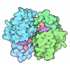
| Title | D. melanogaster Augmin TII N-clamp (GST-fusion) bound to a microtubule, well-defined subset of particles | ||||||||||||||||||||||||||||||
 Components Components |
| ||||||||||||||||||||||||||||||
 Keywords Keywords | CELL CYCLE / augmin / N-clamp / TII / microtubule / branching / nucleation / HAUS / HAUS augmin-like complex subunit / HAUS6 / HAUS7 / HAUS2 / HAUS8 / Dgt6 / Msd5 / Dgt4 / Msd1 / Drosophila melanogaster / complex / CH domain / calponin homology domain / MAP / cell division | ||||||||||||||||||||||||||||||
| Function / homology |  Function and homology information Function and homology informationmitotic spindle-templated microtubule nucleation / HAUS complex / mitotic spindle microtubule / motile cilium / regulation of mitotic nuclear division / glutathione transferase / centrosome duplication / glutathione transferase activity / mitotic spindle assembly / glutathione metabolic process ...mitotic spindle-templated microtubule nucleation / HAUS complex / mitotic spindle microtubule / motile cilium / regulation of mitotic nuclear division / glutathione transferase / centrosome duplication / glutathione transferase activity / mitotic spindle assembly / glutathione metabolic process / bioluminescence / mitotic spindle organization / generation of precursor metabolites and energy / kinetochore / structural constituent of cytoskeleton / microtubule cytoskeleton organization / spindle / neuron migration / spindle pole / mitotic cell cycle / protein-macromolecule adaptor activity / microtubule binding / Hydrolases; Acting on acid anhydrides; Acting on GTP to facilitate cellular and subcellular movement / microtubule / hydrolase activity / cell division / GTPase activity / centrosome / GTP binding / metal ion binding / cytoplasm Similarity search - Function | ||||||||||||||||||||||||||||||
| Biological species |     | ||||||||||||||||||||||||||||||
| Method | ELECTRON MICROSCOPY / single particle reconstruction / cryo EM / Resolution: 4.9 Å | ||||||||||||||||||||||||||||||
 Authors Authors | Wuertz, M. / Vermeulen, B.J.A. / Tonon, G. / Pfeffer, S. | ||||||||||||||||||||||||||||||
| Funding support |  Germany, 9items Germany, 9items
| ||||||||||||||||||||||||||||||
 Citation Citation | Journal: Nat Commun / Year: 2025 Title: Conserved function of the HAUS6 calponin homology domain in anchoring augmin for microtubule branching. Authors: Martin Würtz / Giulia Tonon / Bram J A Vermeulen / Maja Zezlina / Qi Gao / Annett Neuner / Angelika Seidl / Melanie König / Maximilian Harkenthal / Sebastian Eustermann / Sylvia Erhardt / ...Authors: Martin Würtz / Giulia Tonon / Bram J A Vermeulen / Maja Zezlina / Qi Gao / Annett Neuner / Angelika Seidl / Melanie König / Maximilian Harkenthal / Sebastian Eustermann / Sylvia Erhardt / Fabio Lolicato / Elmar Schiebel / Stefan Pfeffer /   Abstract: Branching microtubule nucleation is a key mechanism for mitotic and meiotic spindle assembly and requires the hetero-octameric augmin complex. Augmin recruits the major microtubule nucleator, the γ- ...Branching microtubule nucleation is a key mechanism for mitotic and meiotic spindle assembly and requires the hetero-octameric augmin complex. Augmin recruits the major microtubule nucleator, the γ-tubulin ring complex, to pre-existing microtubules to direct the formation of new microtubules in a defined orientation. Although recent structural work has provided key insights into the structural organization of augmin, molecular details of its interaction with microtubules remain elusive. Here, we identify the minimal conserved microtubule-binding unit of augmin across species and demonstrate that stable microtubule anchoring is predominantly mediated via the calponin homology (CH) domain in Dgt6/HAUS6. Comparative sequence and functional analyses in vitro and in vivo reveal a highly conserved functional role of the HAUS6 CH domain in microtubule binding. Using cryo-electron microscopy and molecular dynamics simulations in combination with AlphaFold structure predictions, we show that the D. melanogaster Dgt6/HAUS6 CH domain binds microtubules at the inter-protofilament groove between two adjacent β-tubulin subunits and thereby orients augmin on microtubules. Altogether, our findings reveal how augmin binds microtubules to pre-determine the branching angle during microtubule nucleation and facilitate the rapid assembly of complex microtubule networks. | ||||||||||||||||||||||||||||||
| History |
|
- Structure visualization
| Structure viewer | Molecule:  Molmil Molmil Jmol/JSmol Jmol/JSmol |
|---|
- Downloads & links
- Download
| PDBx/mmCIF format |  9rpd.cif.gz 9rpd.cif.gz | 873.8 KB | Display |  PDBx/mmCIF format PDBx/mmCIF format |
|---|---|---|---|---|
| PDB format |  pdb9rpd.ent.gz pdb9rpd.ent.gz | 718.5 KB | Display |  PDB format PDB format |
| PDBx/mmJSON format |  9rpd.json.gz 9rpd.json.gz | Tree view |  PDBx/mmJSON format PDBx/mmJSON format | |
| Others |  Other downloads Other downloads |
-Validation report
| Summary document |  9rpd_validation.pdf.gz 9rpd_validation.pdf.gz | 1.3 MB | Display |  wwPDB validaton report wwPDB validaton report |
|---|---|---|---|---|
| Full document |  9rpd_full_validation.pdf.gz 9rpd_full_validation.pdf.gz | 1.3 MB | Display | |
| Data in XML |  9rpd_validation.xml.gz 9rpd_validation.xml.gz | 88.6 KB | Display | |
| Data in CIF |  9rpd_validation.cif.gz 9rpd_validation.cif.gz | 128.6 KB | Display | |
| Arichive directory |  https://data.pdbj.org/pub/pdb/validation_reports/rp/9rpd https://data.pdbj.org/pub/pdb/validation_reports/rp/9rpd ftp://data.pdbj.org/pub/pdb/validation_reports/rp/9rpd ftp://data.pdbj.org/pub/pdb/validation_reports/rp/9rpd | HTTPS FTP |
-Related structure data
| Related structure data | M: map data used to model this data |
|---|---|
| Similar structure data | Similarity search - Function & homology  F&H Search F&H Search |
- Links
Links
- Assembly
| Deposited unit | 
|
|---|---|
| 1 |
|
- Components
| #1: Protein | Mass: 12246.828 Da / Num. of mol.: 1 Source method: isolated from a genetically manipulated source Source: (gene. exp.)   | ||||
|---|---|---|---|---|---|
| #2: Protein | Mass: 32382.451 Da / Num. of mol.: 1 Source method: isolated from a genetically manipulated source Source: (gene. exp.)   | ||||
| #3: Protein | Mass: 76561.094 Da / Num. of mol.: 1 Source method: isolated from a genetically manipulated source Source: (gene. exp.)    Gene: msd5, dgt7, CG2213, GFP / Production host:  References: UniProt: Q9W0G6, UniProt: P42212, UniProt: P08515, glutathione transferase | ||||
| #4: Protein | Mass: 49907.770 Da / Num. of mol.: 2 / Source method: isolated from a natural source / Source: (natural)  #5: Protein | Mass: 50121.266 Da / Num. of mol.: 4 / Source method: isolated from a natural source / Source: (natural)  Has protein modification | N | |
-Experimental details
-Experiment
| Experiment | Method: ELECTRON MICROSCOPY |
|---|---|
| EM experiment | Aggregation state: HELICAL ARRAY / 3D reconstruction method: single particle reconstruction |
- Sample preparation
| Component |
| ||||||||||||||||||||||||
|---|---|---|---|---|---|---|---|---|---|---|---|---|---|---|---|---|---|---|---|---|---|---|---|---|---|
| Molecular weight |
| ||||||||||||||||||||||||
| Source (natural) |
| ||||||||||||||||||||||||
| Source (recombinant) |
| ||||||||||||||||||||||||
| Buffer solution | pH: 6.8 | ||||||||||||||||||||||||
| Specimen | Embedding applied: NO / Shadowing applied: NO / Staining applied: NO / Vitrification applied: YES | ||||||||||||||||||||||||
| Vitrification | Cryogen name: ETHANE |
- Electron microscopy imaging
| Experimental equipment |  Model: Titan Krios / Image courtesy: FEI Company |
|---|---|
| Microscopy | Model: TFS KRIOS |
| Electron gun | Electron source:  FIELD EMISSION GUN / Accelerating voltage: 300 kV / Illumination mode: FLOOD BEAM FIELD EMISSION GUN / Accelerating voltage: 300 kV / Illumination mode: FLOOD BEAM |
| Electron lens | Mode: BRIGHT FIELD / Nominal defocus max: 3000 nm / Nominal defocus min: 1000 nm |
| Image recording | Electron dose: 40.4 e/Å2 / Film or detector model: GATAN K3 BIOQUANTUM (6k x 4k) |
- Processing
| EM software |
| ||||||||||||
|---|---|---|---|---|---|---|---|---|---|---|---|---|---|
| CTF correction | Type: PHASE FLIPPING AND AMPLITUDE CORRECTION | ||||||||||||
| 3D reconstruction | Resolution: 4.9 Å / Resolution method: FSC 0.143 CUT-OFF / Num. of particles: 117870 / Symmetry type: POINT | ||||||||||||
| Atomic model building | Protocol: RIGID BODY FIT |
 Movie
Movie Controller


 PDBj
PDBj


