Movie
Movie Controller

[English] 日本語
 Yorodumi
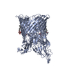
- PDB-9rhr: FusA (ferredoxin receptor from Pectobacterium atrosepticum) in th... -
+ Open data

- Basic information
| Entry | Database: PDB / ID: 9rhr | |||||||||||||||||||||||||||||||||
|---|---|---|---|---|---|---|---|---|---|---|---|---|---|---|---|---|---|---|---|---|---|---|---|---|---|---|---|---|---|---|---|---|---|---|
| Title | FusA (ferredoxin receptor from Pectobacterium atrosepticum) in the presence of Ra-LPS | |||||||||||||||||||||||||||||||||
 Components Components | Ferredoxin receptor | |||||||||||||||||||||||||||||||||
 Keywords Keywords | MEMBRANE PROTEIN / outer membrane protein / transmembrane beta-barrel / tonb-dependent transporter | |||||||||||||||||||||||||||||||||
| Function / homology | TonB-dependent receptor, plug domain superfamily / TonB-dependent receptor, plug domain / TonB-dependent Receptor Plug Domain / TonB-dependent receptor-like, beta-barrel domain superfamily / cell outer membrane / TonB-dependent receptor plug domain-containing protein Function and homology information Function and homology information | |||||||||||||||||||||||||||||||||
| Biological species |  Pectobacterium atrosepticum SCRI1043 (bacteria) Pectobacterium atrosepticum SCRI1043 (bacteria) | |||||||||||||||||||||||||||||||||
| Method | ELECTRON MICROSCOPY / single particle reconstruction / cryo EM / Resolution: 2.4 Å | |||||||||||||||||||||||||||||||||
 Authors Authors | Machin, J.M. / Ranson, N.A. | |||||||||||||||||||||||||||||||||
| Funding support |  United Kingdom, 2items United Kingdom, 2items
| |||||||||||||||||||||||||||||||||
 Citation Citation | Journal: J Struct Biol X / Year: 2025 Title: The structure of the bacterial outer membrane transporter FusA enabled by addition of the native lipid lipopolysaccharide. Authors: Jonathan M Machin / Khedidja Mosbahi / Dheeraj Prakaash / Sheena E Radford / Daniel Walker / Antreas C Kalli / Neil A Ranson / Abstract: Lipopolysaccharide (LPS) is a glycolipid found uniquely in the outer membrane of diderm bacteria, formed of 4-7 acyl chains covalently linked to an extended polysaccharide chain. While a few examples ...Lipopolysaccharide (LPS) is a glycolipid found uniquely in the outer membrane of diderm bacteria, formed of 4-7 acyl chains covalently linked to an extended polysaccharide chain. While a few examples of the interaction between LPS and outer membrane proteins (OMPs) have been structurally characterised, either experimentally or computationally, the precise nature of LPS-OMP interactions and their functional consequences remains unclear. Here, we show that the addition of LPS facilitated cryoEM structure determination of FusA, a 100 kDa TonB-dependent outer membrane transporter from . A 2.8 Å structure combined with molecular dynamics of FusA with different LPS models reveals LPS binding sites with a strong LPS interaction site located adjacent to the β-seam region of the FusA β-barrel. The requirement of lipid binding for successful structure determination indicates a stabilisation of the protein, which in turn suggests a potential method for solving other, small OMPs and membrane proteins. Further, it hints at how LPS may mediate protein conformation and thus how LPS and OMPs can work in concert to maintain a structural and functional OM. | |||||||||||||||||||||||||||||||||
| History |
|
- Structure visualization
| Structure viewer | Molecule: MolmilJmol/JSmol |
|---|
- Downloads & links
-Download
| PDBx/mmCIF format | 9rhr.cif.gz | 290.2 KB | Display | PDBx/mmCIF format |
|---|---|---|---|---|
| PDB format | pdb9rhr.ent.gz | Display | PDB format | |
| PDBx/mmJSON format | 9rhr.json.gz | Tree view | PDBx/mmJSON format | |
| Others |  Other downloads Other downloads |
-Validation report
| Arichive directory | https://data.pdbj.org/pub/pdb/validation_reports/rh/9rhrftp://data.pdbj.org/pub/pdb/validation_reports/rh/9rhr | HTTPS FTP |
|---|
-Related structure data
| Related structure data |  53974MC M: map data used to model this data C: citing same article ( |
|---|---|
| Similar structure data |
-Links
 PDBj
PDBj- Assembly
| Deposited unit | 
|
|---|---|
| 1 |
|
-Components
| #1: Protein | Mass: 97786.562 Da / Num. of mol.: 1 Source method: isolated from a genetically manipulated source Source: (gene. exp.) Pectobacterium atrosepticum SCRI1043 (bacteria)Gene: ECA0878 / Production host: |
|---|---|
| Has protein modification | Y |
-Experimental details
-Experiment
| Experiment | Method: ELECTRON MICROSCOPY |
|---|---|
| EM experiment | Aggregation state: PARTICLE / 3D reconstruction method: single particle reconstruction |
- Sample preparation
| Component | Name: FusA protein in complex with Ra-LPS lipid solubilised in LDAO micelles Type: COMPLEX Details: FusA protein in complex with Ra-LPS lipid solubilised in LDAO micelles. Ra-LPS added into micelles following purification Entity ID: all / Source: RECOMBINANT |
|---|---|
| Molecular weight | Experimental value: NO |
| Source (natural) | Organism: Pectobacterium atrosepticum SCRI1043 (bacteria) |
| Source (recombinant) | Organism: |
| Buffer solution | pH: 8 / Details: 0.2% LDAO, 20 mM Tris-Cl (pH 8.0), 150 mM NaCl |
| Specimen | Conc.: 10 mg/ml / Embedding applied: NO / Shadowing applied: NO / Staining applied: NO / Vitrification applied: YES |
| Specimen support | Grid material: COPPER / Grid type: Quantifoil R1.2/1.3 |
| Vitrification | Instrument: FEI VITROBOT MARK IV / Cryogen name: ETHANE / Humidity: 100 % / Chamber temperature: 4 K |
- Electron microscopy imaging
| Experimental equipment |  Model: Titan Krios / Image courtesy: FEI Company |
|---|---|
| Microscopy | Model: TFS KRIOS |
| Electron gun | Electron source:  FIELD EMISSION GUN / Accelerating voltage: 300 kV / Illumination mode: FLOOD BEAM FIELD EMISSION GUN / Accelerating voltage: 300 kV / Illumination mode: FLOOD BEAM |
| Electron lens | Mode: BRIGHT FIELD / Nominal defocus max: 30000 nm / Nominal defocus min: 9000 nm / C2 aperture diameter: 70 µm |
| Image recording | Electron dose: 47.1 e/Å2 / Film or detector model: TFS FALCON 4i (4k x 4k) |
| EM imaging optics | Energyfilter name: TFS Selectris / Energyfilter slit width: 5 eV |
- Processing
| EM software |
| ||||||||||||||||||||||||||||||||||||||||
|---|---|---|---|---|---|---|---|---|---|---|---|---|---|---|---|---|---|---|---|---|---|---|---|---|---|---|---|---|---|---|---|---|---|---|---|---|---|---|---|---|---|
| CTF correction | Type: PHASE FLIPPING AND AMPLITUDE CORRECTION | ||||||||||||||||||||||||||||||||||||||||
| Particle selection | Num. of particles selected: 754778 | ||||||||||||||||||||||||||||||||||||||||
| 3D reconstruction | Resolution: 2.4 Å / Resolution method: FSC 0.143 CUT-OFF / Num. of particles: 84061 / Symmetry type: POINT | ||||||||||||||||||||||||||||||||||||||||
| Atomic model building | Protocol: FLEXIBLE FIT / Space: REAL / Details: Isolde and phenix real-space refine | ||||||||||||||||||||||||||||||||||||||||
| Atomic model building | PDB-ID: 4ZGV Accession code: 4ZGV / Source name: PDB / Type: experimental model |