 Movie
Movie Controller


+ Open data

- Basic information
| Entry | Database: PDB / ID: 9rbu | |||||||||||||||||||||||||||
|---|---|---|---|---|---|---|---|---|---|---|---|---|---|---|---|---|---|---|---|---|---|---|---|---|---|---|---|---|
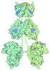
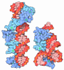
| Title | Cryo-ET structure of full-length membrane-bound EHD2 complex | |||||||||||||||||||||||||||
 Components Components | EH domain-containing protein 2 | |||||||||||||||||||||||||||
 Keywords Keywords | STRUCTURAL PROTEIN / Eps15-homology domain-containing proteins (EHDs) / dynamin-related ATPases / membrane remodeling / human myotubes / membrane repair process / caveolae / EHD2 / lipid homeostasis / plasma membrane invaginations / oligomer / nucleotide binding / EHD2-dependent caveolae stabilization / GTPase (G-) domain / ATPase activity / liposome tubulation | |||||||||||||||||||||||||||
| Function / homology |  Function and homology information Function and homology informationpositive regulation of endocytic recycling / plasma membrane tubulation / Factors involved in megakaryocyte development and platelet production / endocytic recycling / cortical actin cytoskeleton organization / positive regulation of myoblast fusion / endocytic vesicle / cilium assembly / protein localization to plasma membrane / endocytosis ...positive regulation of endocytic recycling / plasma membrane tubulation / Factors involved in megakaryocyte development and platelet production / endocytic recycling / cortical actin cytoskeleton organization / positive regulation of myoblast fusion / endocytic vesicle / cilium assembly / protein localization to plasma membrane / endocytosis / caveola / recycling endosome membrane / hydrolase activity / early endosome / protein-macromolecule adaptor activity / protein domain specific binding / calcium ion binding / GTP binding / perinuclear region of cytoplasm / ATP binding / membrane / identical protein binding / plasma membrane / cytosol / cytoplasm Similarity search - Function | |||||||||||||||||||||||||||
| Biological species |   Mus musculus (house mouse) Mus musculus (house mouse) | |||||||||||||||||||||||||||
| Method | ELECTRON MICROSCOPY / subtomogram averaging / cryo EM / Resolution: 6.7 Å | |||||||||||||||||||||||||||
 Authors Authors | Vazquez-Sarandeses, E. / Mikirtumov, V. / Noel, J. / Kudryashev, M. / Daumke, O. | |||||||||||||||||||||||||||
| Funding support |  Germany, 1items Germany, 1items
| |||||||||||||||||||||||||||
 Citation Citation | Journal: Nat Commun / Year: 2026 Title: Structures of EHD2 filaments on curved membranes provide a model for caveolar neck stabilization. Authors: Elena Vázquez-Sarandeses / Vasilii Mikirtumov / Jeffrey K Noel / Mikhail Kudryashev / Oliver Daumke / Abstract: Caveolae are flask-shaped invaginations of the plasma membrane serving critical functions in mechano-protection and signal transduction. Caveolar dynamics, such as caveolar movement within the plasma ...Caveolae are flask-shaped invaginations of the plasma membrane serving critical functions in mechano-protection and signal transduction. Caveolar dynamics, such as caveolar movement within the plasma membrane or endocytosis, relies on precise shaping of the highly curved caveolar necks. The dynamin-like EHD2 ATPase is proposed to oligomerize around the caveolar neck, but its detailed molecular action is poorly understood. Here, we employ cryo-electron tomography to elucidate structures of ring-like EHD2 filaments on tubulated liposomes. EHD2 forms highly curved membrane scaffolds which stabilize a tubular membrane geometry with undulations along the tube's axis, resembling caveolar neck architecture. An amino-terminal sequence facilitates this geometry by acting as a spacer between adjacent filaments. Moreover, in endothelial cells lacking EHD2, caveolar necks become narrower and more elongated. Our structural work provides the molecular framework for understanding EHD2 scaffold formation and its cellular function in caveolar dynamics. #1: Journal: biorxiv.org / Year: 2025Title: Structures of EHD2 filaments on curved membranes provides a model for caveolar neck stabilization Authors: Vazquez-Sarandeses, E. / Mikirtumov, V. / Noel, J. / Kudryashev, M. / Daumke, O. | |||||||||||||||||||||||||||
| History |
|
- Structure visualization
| Structure viewer | Molecule: MolmilJmol/JSmol |
|---|
- Downloads & links
-Download
| PDBx/mmCIF format | 9rbu.cif.gz | 315.9 KB | Display | PDBx/mmCIF format |
|---|---|---|---|---|
| PDB format | pdb9rbu.ent.gz | 243.5 KB | Display | PDB format |
| PDBx/mmJSON format | 9rbu.json.gz | Tree view | PDBx/mmJSON format | |
| Others |  Other downloads Other downloads |
-Validation report
| Arichive directory | https://data.pdbj.org/pub/pdb/validation_reports/rb/9rbuftp://data.pdbj.org/pub/pdb/validation_reports/rb/9rbu | HTTPS FTP |
|---|
-Related structure data
| Related structure data |  53909MC  9rc1C M: map data used to model this data C: citing same article ( |
|---|---|
| Similar structure data |

-Links
 PDBj
PDBj
- Assembly
| Deposited unit | 
|
|---|---|
| 1 |
|
-Components
| #1: Protein | Mass: 61277.418 Da / Num. of mol.: 4 Source method: isolated from a genetically manipulated source Details: Expressed in E. coli (BL21(DE3)-Rosetta2 strain) from a modified pET28 vector as N-terminal His6-tag fusions followed by a PreScission protease cleavage site. Source: (gene. exp.) Mus musculus (house mouse) / Gene: Ehd2 / Production host:   Escherichia coli BL21(DE3) (bacteria) / Variant (production host): -Rosetta2 / References: UniProt: Q8BH64 Escherichia coli BL21(DE3) (bacteria) / Variant (production host): -Rosetta2 / References: UniProt: Q8BH64#2: Chemical | ChemComp-ATP / 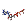 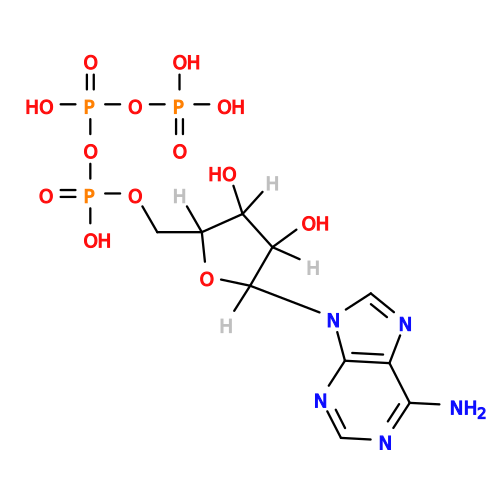 Mass: 507.181 Da / Num. of mol.: 4 / Source method: obtained synthetically / Formula: C10H16N5O13P3 / Feature type: SUBJECT OF INVESTIGATION / Comment: ATP, energy-carrying molecule*YM Mass: 507.181 Da / Num. of mol.: 4 / Source method: obtained synthetically / Formula: C10H16N5O13P3 / Feature type: SUBJECT OF INVESTIGATION / Comment: ATP, energy-carrying molecule*YM#3: Chemical | ChemComp-MG /   Mass: 24.305 Da / Num. of mol.: 4 / Source method: obtained synthetically / Formula: Mg / Feature type: SUBJECT OF INVESTIGATION Mass: 24.305 Da / Num. of mol.: 4 / Source method: obtained synthetically / Formula: Mg / Feature type: SUBJECT OF INVESTIGATIONHas ligand of interest | Y | Has protein modification | N | |
|---|
-Experimental details
-Experiment
| Experiment | Method: ELECTRON MICROSCOPY |
|---|---|
| EM experiment | Aggregation state: FILAMENT / 3D reconstruction method: subtomogram averaging |
- Sample preparation
| Component | Name: EHD2 with ATP and liposomes / Type: ORGANELLE OR CELLULAR COMPONENT / Entity ID: #1 / Source: RECOMBINANT |
|---|---|
| Molecular weight | Experimental value: NO |
| Source (natural) | Organism: Mus musculus (house mouse) |
| Source (recombinant) | Organism: Escherichia coli (E. coli) |
| Buffer solution | pH: 7.5 / Details: 20 mM HEPES/NaOH pH 7.5, 300 mM NaCl, 0.5 mM MgCl2 |
| Specimen | Conc.: 2 mg/ml / Embedding applied: NO / Shadowing applied: NO / Staining applied: NO / Vitrification applied: YES |
| Specimen support | Grid material: COPPER / Grid mesh size: 200 divisions/in. / Grid type: Quantifoil R2/2 |
| Vitrification | Instrument: FEI VITROBOT MARK II / Cryogen name: ETHANE / Humidity: 95 % / Chamber temperature: 298 K |
- Electron microscopy imaging
| Experimental equipment |  Model: Titan Krios / Image courtesy: FEI Company |
|---|---|
| Microscopy | Model: TFS KRIOS |
| Electron gun | Electron source:  FIELD EMISSION GUN / Accelerating voltage: 300 kV / Illumination mode: FLOOD BEAM FIELD EMISSION GUN / Accelerating voltage: 300 kV / Illumination mode: FLOOD BEAM |
| Electron lens | Mode: BRIGHT FIELD / Nominal magnification: 42000 X / Calibrated magnification: 42000 X / Nominal defocus max: 7000 nm / Nominal defocus min: 2000 nm / Calibrated defocus min: 2000 nm / Calibrated defocus max: 7000 nm / Cs: 2.7 mm / Alignment procedure: COMA FREE |
| Specimen holder | Cryogen: NITROGEN / Specimen holder model: FEI TITAN KRIOS AUTOGRID HOLDER |
| Image recording | Electron dose: 2 e/Å2 / Avg electron dose per subtomogram: 100 e/Å2 / Film or detector model: GATAN K3 (6k x 4k) / Num. of real images: 1 |
| EM imaging optics | Energyfilter name: GIF Bioquantum / Energyfilter slit width: 20 eV |
- Processing
| EM software |
| ||||||||||||||||||||||||||||||||||||
|---|---|---|---|---|---|---|---|---|---|---|---|---|---|---|---|---|---|---|---|---|---|---|---|---|---|---|---|---|---|---|---|---|---|---|---|---|---|
| CTF correction | Type: PHASE FLIPPING AND AMPLITUDE CORRECTION | ||||||||||||||||||||||||||||||||||||
| Symmetry | Point symmetry: C2 (2 fold cyclic) | ||||||||||||||||||||||||||||||||||||
| 3D reconstruction | Resolution: 6.7 Å / Resolution method: FSC 0.143 CUT-OFF / Num. of particles: 75439 / Algorithm: FOURIER SPACE / Num. of class averages: 1 / Symmetry type: POINT | ||||||||||||||||||||||||||||||||||||
| EM volume selection | Method: supersampling filament surface / Num. of tomograms: 61 / Num. of volumes extracted: 14491 / Reference model: not applicable | ||||||||||||||||||||||||||||||||||||
| Atomic model building | Protocol: FLEXIBLE FIT / Details: MDfit (Whitford et al., 2011) | ||||||||||||||||||||||||||||||||||||
| Atomic model building | PDB-ID: 4cid Accession code: 4cid / Source name: PDB / Type: experimental model |

