[English] 日本語
Yorodumi
- EMDB-53909: Cryo-ET structure of full-length membrane-bound EHD2 complex -

+
Open data


ID or keywords:

Loading...

-
Basic information

Entry
Database: EMDB / ID: EMD-53909
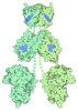
TitleCryo-ET structure of full-length membrane-bound EHD2 complex
Map dataSharpened with an ad-hoc B-factor of -200.
Sample
  • Organelle or cellular component: EHD2 with ATP and liposomes
    • Protein or peptide: EH domain-containing protein 2
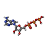
  • Ligand: ADENOSINE-5'-TRIPHOSPHATE
  • Ligand: MAGNESIUM ION
KeywordsEps15-homology domain-containing proteins (EHDs) / dynamin-related ATPases / membrane remodeling / human myotubes / membrane repair process / caveolae / EHD2 / lipid homeostasis / plasma membrane invaginations / oligomer / nucleotide binding / EHD2-dependent caveolae stabilization / GTPase (G-) domain / ATPase activity / liposome tubulation / STRUCTURAL PROTEIN
Function / homology
Function and homology information


positive regulation of endocytic recycling / plasma membrane tubulation / Factors involved in megakaryocyte development and platelet production / endocytic recycling / cortical actin cytoskeleton organization / positive regulation of myoblast fusion / cilium assembly / endocytic vesicle / protein localization to plasma membrane / caveola ...positive regulation of endocytic recycling / plasma membrane tubulation / Factors involved in megakaryocyte development and platelet production / endocytic recycling / cortical actin cytoskeleton organization / positive regulation of myoblast fusion / cilium assembly / endocytic vesicle / protein localization to plasma membrane / caveola / recycling endosome membrane / endocytosis / protein-macromolecule adaptor activity / early endosome / protein domain specific binding / hydrolase activity / calcium ion binding / GTP binding / perinuclear region of cytoplasm / ATP binding / membrane / identical protein binding / plasma membrane / cytoplasm / cytosol
Similarity search - Function
EH domain-containing protein, N-terminal / Domain of unknown function DUF5600 / N-terminal EH-domain containing protein / Domain of unknown function (DUF5600) / EH domain / EH domain profile. / Eps15 homology domain / EH domain / Dynamin-type guanine nucleotide-binding (G) domain / Dynamin-type guanine nucleotide-binding (G) domain profile. ...EH domain-containing protein, N-terminal / Domain of unknown function DUF5600 / N-terminal EH-domain containing protein / Domain of unknown function (DUF5600) / EH domain / EH domain profile. / Eps15 homology domain / EH domain / Dynamin-type guanine nucleotide-binding (G) domain / Dynamin-type guanine nucleotide-binding (G) domain profile. / Dynamin, N-terminal / Dynamin family / EF-Hand 1, calcium-binding site / EF-hand calcium-binding domain. / EF-hand calcium-binding domain profile. / EF-hand domain / EF-hand domain pair / P-loop containing nucleoside triphosphate hydrolase
Similarity search - Domain/homology
EH domain-containing protein 2
Similarity search - Component
Biological speciesMus musculus (house mouse)
Methodsubtomogram averaging / cryo EM / Resolution: 6.7 Å
AuthorsVazquez-Sarandeses E / Mikirtumov V / Noel J / Kudryashev M / Daumke O
Funding support Germany, 1 items
OrganizationGrant numberCountry
Helmholtz Association Germany
CitationJournal: biorxiv.org / Year: 2025
Title: Structures of EHD2 filaments on curved membranes provides a model for caveolar neck stabilization
Authors: Vazquez-Sarandeses E / Mikirtumov V / Noel J / Kudryashev M / Daumke O
History
DepositionMay 27, 2025-
Header (metadata) releaseJul 8, 2026-
Map releaseJul 8, 2026-
UpdateJul 8, 2026-
Current statusJul 8, 2026Processing site: PDBe / Status: Released

-
Structure visualization

Supplemental images

Downloads & links

-
Map

FileReleased
AnnotationSharpened with an ad-hoc B-factor of -200.
Projections & slices

Image control

Size
Brightness
Contrast
Others
AxesZ (Sec.)Y (Row.)X (Col.)
2.14 Å/pix.
x 192 pix.
= 410.496 Å
2.14 Å/pix.
x 192 pix.
= 410.496 Å
2.14 Å/pix.
x 192 pix.
= 410.496 Å

Surface

Projections

Slices (1/3)

Slices (1/2)

Slices (2/3)

Images are generated by Spider.

Voxel sizeX=Y=Z: 2.138 Å
Density
Contour LevelBy AUTHOR: 0.0146
Minimum - Maximum-0.03004479 - 0.13712315
Average (Standard dev.)0.00014659994 (±0.002177657)
SymmetrySpace group: 1
Details

EMDB XML:

Map geometry
Axis orderXYZ
Origin000
Dimensions192192192
Spacing192192192
CellA=B=C: 410.496 Å
α=β=γ: 90.0 °

-
Supplemental data

-
Mask #1

Fileemd_53909_msk_1.map
Projections & Slices
AxesZYX

Projections

Slices (1/2)
Density Histograms

-
Half map: #2

Fileemd_53909_half_map_1.map
Projections & Slices
AxesZYX

Projections

Slices (1/2)
Density Histograms

-
Half map: #1

Fileemd_53909_half_map_2.map
Projections & Slices
AxesZYX

Projections

Slices (1/2)
Density Histograms

-
Sample components

-
Entire : EHD2 with ATP and liposomes

EntireName: EHD2 with ATP and liposomes
Components
  • Organelle or cellular component: EHD2 with ATP and liposomes
    • Protein or peptide: EH domain-containing protein 2
  • Ligand: ADENOSINE-5'-TRIPHOSPHATE
  • Ligand: MAGNESIUM ION

-
Supramolecule #1: EHD2 with ATP and liposomes

SupramoleculeName: EHD2 with ATP and liposomes / type: organelle_or_cellular_component / ID: 1 / Parent: 0 / Macromolecule list: #1
Source (natural)Organism: Mus musculus (house mouse)

-
Macromolecule #1: EH domain-containing protein 2

MacromoleculeName: EH domain-containing protein 2 / type: protein_or_peptide / ID: 1
Details: Expressed in E. coli (BL21(DE3)-Rosetta2 strain) from a modified pET28 vector as N-terminal His6-tag fusions followed by a PreScission protease cleavage site.
Number of copies: 4 / Enantiomer: LEVO
Source (natural)Organism: Mus musculus (house mouse)
Molecular weightTheoretical: 61.277418 KDa
Recombinant expressionOrganism: Escherichia coli BL21(DE3) (bacteria)
SequenceString: MFSWMKKGGA RGQRPEAIRT VTSSLKELYR TKLLPLEEHY RFGSFHSPAL EDADFDGKPM VLVAGQYSTG KTSFIQYLLE QEVPGSRVG PEPTTDCFVA VMHGETEGTV PGNALVVDPE KPFRKLNPFG NTFLNRFMCA QLPNQVLESI SIIDTPGILS G AKQRVSRG ...String:
MFSWMKKGGA RGQRPEAIRT VTSSLKELYR TKLLPLEEHY RFGSFHSPAL EDADFDGKPM VLVAGQYSTG KTSFIQYLLE QEVPGSRVG PEPTTDCFVA VMHGETEGTV PGNALVVDPE KPFRKLNPFG NTFLNRFMCA QLPNQVLESI SIIDTPGILS G AKQRVSRG YDFPAVLRWF AERVDLIILL FDAHKLEISD EFSEAIGALR GHEDKIRVVL NKADMVETQQ LMRVYGALMW AL GKVVGTP EVLRVYIGSF WSQPLLVPDN RRLFELEEQD LFRDIQGLPR HAALRKLNDL VKRARLVRVH AYIISYLKKE MPT VFGKEN KKKQLILKLP VIFAKIQLEH HISPGDFPDC QKMQELLMAH DFTKFHSLKP KLLEALDDML AQDIAKLMPL LRQE ELESV EAGVQGGAFE GTRMGPFVER GPDEAIEDGE EGSEDDAEWV VTKDKSKYDE IFYNLAPADG KLSGSKAKTW MVGTK LPNS VLGRIWKLSD VDRDGMLDDE EFALASHLIE AKLEGHGLPT NLPRRLVPPS KRRQKGSAE

UniProtKB: EH domain-containing protein 2

-
Macromolecule #2: ADENOSINE-5'-TRIPHOSPHATE

MacromoleculeName: ADENOSINE-5'-TRIPHOSPHATE / type: ligand / ID: 2 / Number of copies: 4 / Formula: ATP
Molecular weightTheoretical: 507.181 Da
Chemical component information

ChemComp-ATP:
ADENOSINE-5'-TRIPHOSPHATE / ATP, energy-carrying molecule*YM

-
Macromolecule #3: MAGNESIUM ION

MacromoleculeName: MAGNESIUM ION / type: ligand / ID: 3 / Number of copies: 4 / Formula: MG
Molecular weightTheoretical: 24.305 Da

-
Experimental details

-
Structure determination

Methodcryo EM
Processingsubtomogram averaging
Aggregation statefilament

-
Sample preparation

Concentration2 mg/mL
BufferpH: 7.5 / Details: 20 mM HEPES/NaOH pH 7.5, 300 mM NaCl, 0.5 mM MgCl2
GridModel: Quantifoil R2/2 / Material: COPPER / Mesh: 200 / Support film - Material: CARBON / Support film - topology: HOLEY / Pretreatment - Type: GLOW DISCHARGE / Pretreatment - Time: 90 sec.
VitrificationCryogen name: ETHANE / Chamber humidity: 95 % / Chamber temperature: 298 K / Instrument: FEI VITROBOT MARK II

-
Electron microscopy

MicroscopeTFS KRIOS
Specialist opticsEnergy filter - Name: GIF Bioquantum / Energy filter - Slit width: 20 eV
Image recordingFilm or detector model: GATAN K3 (6k x 4k) / Number real images: 1 / Average electron dose: 2.0 e/Å2
Electron beamAcceleration voltage: 300 kV / Electron source: FIELD EMISSION GUN
Electron opticsCalibrated defocus max: 7.0 µm / Calibrated defocus min: 2.0 µm / Calibrated magnification: 42000 / Illumination mode: FLOOD BEAM / Imaging mode: BRIGHT FIELD / Cs: 2.7 mm / Nominal defocus max: 7.0 µm / Nominal defocus min: 2.0 µm / Nominal magnification: 42000
Sample stageSpecimen holder model: FEI TITAN KRIOS AUTOGRID HOLDER / Cooling holder cryogen: NITROGEN
Experimental equipment
Model: Titan Krios / Image courtesy: FEI Company

+
Image processing

Final reconstructionNumber classes used: 1 / Applied symmetry - Point group: C2 (2 fold cyclic) / Algorithm: FOURIER SPACE / Resolution.type: BY AUTHOR / Resolution: 6.7 Å / Resolution method: FSC 0.143 CUT-OFF / Software - Name: RELION (ver. 4) / Number subtomograms used: 75439
ExtractionNumber tomograms: 61 / Number images used: 14491 / Reference model: not applicable / Method: supersampling filament surface / Software - Name: Dynamo
CTF correctionSoftware: (Name: Gctf, RELION (ver. 4)) / Type: PHASE FLIPPING AND AMPLITUDE CORRECTION
Final angle assignmentType: MAXIMUM LIKELIHOOD / Software: (Name: RELION (ver. 4), cryoSPARC)
FSC plot (resolution estimation)

-
Atomic model buiding 1

Initial modelPDB ID:

Chain - Source name: PDB / Chain - Initial model type: experimental model
DetailsMDfit (Whitford et al., 2011)
RefinementProtocol: FLEXIBLE FIT
Output model

PDB-9rbu:
Cryo-ET structure of full-length membrane-bound EHD2 complex

+
About Yorodumi

-
News

-
Feb 9, 2022. New format data for meta-information of EMDB entries

New format data for meta-information of EMDB entries

  • Version 3 of the EMDB header file is now the official format.
  • The previous official version 1.9 will be removed from the archive.

Related info.:EMDB header

External links:wwPDB to switch to version 3 of the EMDB data model

-
Aug 12, 2020. Covid-19 info

Covid-19 info

URL: https://pdbj.org/emnavi/covid19.php

New page: Covid-19 featured information page in EM Navigator.

Related info.:Covid-19 info / Mar 5, 2020. Novel coronavirus structure data

+
Mar 5, 2020. Novel coronavirus structure data

Novel coronavirus structure data

Related info.:Yorodumi Speices / Aug 12, 2020. Covid-19 info

External links:COVID-19 featured content - PDBj / Molecule of the Month (242):Coronavirus Proteases

+
Jan 31, 2019. EMDB accession codes are about to change! (news from PDBe EMDB page)

EMDB accession codes are about to change! (news from PDBe EMDB page)

  • The allocation of 4 digits for EMDB accession codes will soon come to an end. Whilst these codes will remain in use, new EMDB accession codes will include an additional digit and will expand incrementally as the available range of codes is exhausted. The current 4-digit format prefixed with “EMD-” (i.e. EMD-XXXX) will advance to a 5-digit format (i.e. EMD-XXXXX), and so on. It is currently estimated that the 4-digit codes will be depleted around Spring 2019, at which point the 5-digit format will come into force.
  • The EM Navigator/Yorodumi systems omit the EMD- prefix.

Related info.:Q: What is EMD? / ID/Accession-code notation in Yorodumi/EM Navigator

External links:EMDB Accession Codes are Changing Soon! / Contact to PDBj

+
Jul 12, 2017. Major update of PDB

Major update of PDB

  • wwPDB released updated PDB data conforming to the new PDBx/mmCIF dictionary.
  • This is a major update changing the version number from 4 to 5, and with Remediation, in which all the entries are updated.
  • In this update, many items about electron microscopy experimental information are reorganized (e.g. em_software).
  • Now, EM Navigator and Yorodumi are based on the updated data.

External links:wwPDB Remediation / Enriched Model Files Conforming to OneDep Data Standards Now Available in the PDB FTP Archive

-
Yorodumi

Thousand views of thousand structures

  • Yorodumi is a browser for structure data from EMDB, PDB, SASBDB, etc.
  • This page is also the successor to EM Navigator detail page, and also detail information page/front-end page for Omokage search.
  • The word "yorodu" (or yorozu) is an old Japanese word meaning "ten thousand". "mi" (miru) is to see.

Related info.:EMDB / PDB / SASBDB / Comparison of 3 databanks / Yorodumi Search / Aug 31, 2016. New EM Navigator & Yorodumi / Yorodumi Papers / Jmol/JSmol / Function and homology information / Changes in new EM Navigator and Yorodumi

Read more