 Movie
Movie Controller


+ Open data

- Basic information
| Entry | Database: PDB / ID: 9qua | |||||||||
|---|---|---|---|---|---|---|---|---|---|---|
| Title | apPol-DNA-nucleotide complex (ternary 1) | |||||||||
 Components Components |
| |||||||||
 Keywords Keywords | REPLICATION / DNA polymerase | |||||||||
| Function / homology |  Function and homology information Function and homology information3'-5' exonuclease activity / DNA-templated DNA replication / single-stranded DNA binding / 5'-3' DNA helicase activity / DNA-directed DNA polymerase activity / ATP binding Similarity search - Function | |||||||||
| Biological species |  DNA molecule (others) | |||||||||
| Method | ELECTRON MICROSCOPY / single particle reconstruction / cryo EM / Resolution: 4.2 Å | |||||||||
 Authors Authors | Lahiri, I. / Kumari, A. | |||||||||
| Funding support |  India, India,  United Kingdom, 2items United Kingdom, 2items
| |||||||||
 Citation Citation | Journal: Nucleic Acids Res / Year: 2025 Title: Structural basis of multitasking by the apicoplast DNA polymerase from Plasmodium falciparum. Authors: Anamika Kumari / Theodora Enache / Timothy D Craggs / Janice D Pata / Indrajit Lahiri /  Abstract: Plasmodium falciparum is a eukaryotic pathogen responsible for the majority of malaria-related fatalities. Plasmodium belongs to the phylum Apicomplexa and, like most members of this phylum, contains ...Plasmodium falciparum is a eukaryotic pathogen responsible for the majority of malaria-related fatalities. Plasmodium belongs to the phylum Apicomplexa and, like most members of this phylum, contains a non-photosynthetic plastid called the apicoplast. The apicoplast has its own genome, replicated by a dedicated replisome. Unlike other cellular replisomes, the apicoplast replisome uses a single DNA polymerase (apPol). This suggests that apPol can multitask and catalyse both replicative and lesion bypass synthesis. Replicative synthesis relies on a restrictive active site for high accuracy while lesion bypass typically requires an open active site. This raises the question: how does apPol combine the structural features of multiple DNA polymerases in a single protein? Using single-particle electron cryomicroscopy (cryoEM), we have solved the structures of apPol bound to its undamaged DNA and nucleotide substrates in five pre-chemistry conformational states. We found that apPol can accommodate a nascent base pair with the fingers in an open configuration, which might facilitate the lesion bypass activity. In the fingers-open state, we identified a nascent base pair checkpoint that preferentially selects Watson-Crick base pairs, an essential requirement for replicative synthesis. Taken together, these structural features might explain how apPol balances replicative and lesion bypass synthesis. | |||||||||
| History |
|
- Structure visualization
| Structure viewer | Molecule: MolmilJmol/JSmol |
|---|
- Downloads & links
-Download
| PDBx/mmCIF format | 9qua.cif.gz | 151.3 KB | Display | PDBx/mmCIF format |
|---|---|---|---|---|
| PDB format | pdb9qua.ent.gz | 111.2 KB | Display | PDB format |
| PDBx/mmJSON format | 9qua.json.gz | Tree view | PDBx/mmJSON format | |
| Others |  Other downloads Other downloads |
-Validation report
| Summary document | 9qua_validation.pdf.gz | 1.4 MB | Display | wwPDB validaton report |
|---|---|---|---|---|
| Full document | 9qua_full_validation.pdf.gz | 1.4 MB | Display | |
| Data in XML | 9qua_validation.xml.gz | 33.1 KB | Display | |
| Data in CIF | 9qua_validation.cif.gz | 48.6 KB | Display | |
| Arichive directory | https://data.pdbj.org/pub/pdb/validation_reports/qu/9quaftp://data.pdbj.org/pub/pdb/validation_reports/qu/9qua | HTTPS FTP |
-Related structure data
| Related structure data |  53376MC  9qscC  9qu8C  9qujC  9qunC  9qv9C C: citing same article ( M: map data used to model this data |
|---|---|
| Similar structure data |


-Links
 PDBj
PDBj


- Assembly
| Deposited unit | 
|
|---|---|
| 1 |
|
-Components
| #1: Protein | Mass: 76473.484 Da / Num. of mol.: 1 Source method: isolated from a genetically manipulated source Source: (gene. exp.) Gene: PFMALIP_04965 / Production host:  |
|---|---|
| #2: DNA chain | Mass: 7714.971 Da / Num. of mol.: 1 / Source method: obtained synthetically / Details: DNA primer strand / Source: (synth.) DNA molecule (others) |
| #3: DNA chain | Mass: 11023.080 Da / Num. of mol.: 1 / Source method: obtained synthetically / Source: (synth.) DNA molecule (others) |
| #4: Chemical | ChemComp-DGT / 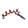  Mass: 507.181 Da / Num. of mol.: 1 / Source method: obtained synthetically / Formula: C10H16N5O13P3 / Feature type: SUBJECT OF INVESTIGATION Mass: 507.181 Da / Num. of mol.: 1 / Source method: obtained synthetically / Formula: C10H16N5O13P3 / Feature type: SUBJECT OF INVESTIGATION |
| Has ligand of interest | Y |
| Has protein modification | N |
-Experimental details
-Experiment
| Experiment | Method: ELECTRON MICROSCOPY |
|---|---|
| EM experiment | Aggregation state: PARTICLE / 3D reconstruction method: single particle reconstruction |
- Sample preparation
| Component | Name: Ternary complex of apicoplast DNA polymerase with DNA and dGTP Type: COMPLEX / Entity ID: #1-#3 / Source: RECOMBINANT |
|---|---|
| Molecular weight | Value: 0.095 MDa / Experimental value: NO |
| Source (natural) | Organism: |
| Source (recombinant) | Organism: |
| Buffer solution | pH: 7.5 |
| Specimen | Embedding applied: NO / Shadowing applied: NO / Staining applied: NO / Vitrification applied: YES |
| Vitrification | Cryogen name: ETHANE |
- Electron microscopy imaging
| Experimental equipment |  Model: Titan Krios / Image courtesy: FEI Company |
|---|---|
| Microscopy | Model: TFS KRIOS |
| Electron gun | Electron source:  FIELD EMISSION GUN / Accelerating voltage: 300 kV / Illumination mode: FLOOD BEAM FIELD EMISSION GUN / Accelerating voltage: 300 kV / Illumination mode: FLOOD BEAM |
| Electron lens | Mode: BRIGHT FIELD / Nominal magnification: 81000 X / Calibrated magnification: 81000 X / Nominal defocus max: 3800 nm / Nominal defocus min: 800 nm / Calibrated defocus min: 800 nm / Calibrated defocus max: 3800 nm / Cs: 2.7 mm / C2 aperture diameter: 50 µm / Alignment procedure: COMA FREE |
| Specimen holder | Cryogen: NITROGEN / Specimen holder model: FEI TITAN KRIOS AUTOGRID HOLDER / Temperature (max): 77 K / Temperature (min): 77 K |
| Image recording | Average exposure time: 1 sec. / Electron dose: 70 e/Å2 / Film or detector model: GATAN K3 BIOCONTINUUM (6k x 4k) / Num. of grids imaged: 1 / Num. of real images: 16189 |
| EM imaging optics | Energyfilter slit width: 20 eV |
- Processing
| EM software |
| ||||||||||||||||||||||||||||||||||||||||
|---|---|---|---|---|---|---|---|---|---|---|---|---|---|---|---|---|---|---|---|---|---|---|---|---|---|---|---|---|---|---|---|---|---|---|---|---|---|---|---|---|---|
| CTF correction | Type: PHASE FLIPPING AND AMPLITUDE CORRECTION | ||||||||||||||||||||||||||||||||||||||||
| Particle selection | Num. of particles selected: 1922870 | ||||||||||||||||||||||||||||||||||||||||
| 3D reconstruction | Resolution: 4.2 Å / Resolution method: FSC 0.143 CUT-OFF / Num. of particles: 9780 / Symmetry type: POINT | ||||||||||||||||||||||||||||||||||||||||
| Atomic model building | Protocol: RIGID BODY FIT | ||||||||||||||||||||||||||||||||||||||||
| Atomic model building | PDB-ID: 5DKT Accession code: 5DKT / Source name: PDB / Type: experimental model | ||||||||||||||||||||||||||||||||||||||||
| Refinement | Highest resolution: 4.2 Å Stereochemistry target values: REAL-SPACE (WEIGHTED MAP SUM AT ATOM CENTERS) | ||||||||||||||||||||||||||||||||||||||||
| Refine LS restraints |
|

