 Movie
Movie Controller

[English] 日本語
 Yorodumi
- PDB-9qr1: Methyl-coenzyme M reductase of ANME-2d Candidatus Methanoperedens... -
+ Open data

- Basic information
| Entry | Database: PDB / ID: 9qr1 | ||||||||||||||||||
|---|---|---|---|---|---|---|---|---|---|---|---|---|---|---|---|---|---|---|---|
| Title | Methyl-coenzyme M reductase of ANME-2d Candidatus Methanoperedens sp. BLZ2 from a bioreactor enrichment culture | ||||||||||||||||||
 Components Components |
| ||||||||||||||||||
 Keywords Keywords | TRANSFERASE / methanotrophy / anaerobic methanotrophic archaea / methane oxidation / ANME / methyl-coenzyme M reductase / MCR / reverse methanogenesis / nickel-dependent enzyme / coenzyme M / coenzyme B / F430 cofactor | ||||||||||||||||||
| Function / homology |  Function and homology information Function and homology informationcoenzyme-B sulfoethylthiotransferase / coenzyme-B sulfoethylthiotransferase activity / methanogenesis / metal ion binding Similarity search - Function | ||||||||||||||||||
| Biological species |  Candidatus Methanoperedens sp. BLZ2 (archaea) Candidatus Methanoperedens sp. BLZ2 (archaea) | ||||||||||||||||||
| Method |  X-RAY DIFFRACTION / SYNCHROTRON / MOLECULAR REPLACEMENT / Resolution: 0.98 Å X-RAY DIFFRACTION / SYNCHROTRON / MOLECULAR REPLACEMENT / Resolution: 0.98 Å | ||||||||||||||||||
 Authors Authors | Mueller, M.-C. / Wagner, T. | ||||||||||||||||||
| Funding support | European Union,  Germany, Germany,  Netherlands, 5items Netherlands, 5items
| ||||||||||||||||||
 Citation Citation | Journal: To Be Published Title: Atomic resolution structures of the key enzyme MCR in anaerobic methanotrophy reveal novel and extensive post-translational modifications. Authors: Mueller, M.-C. / Wissink, M. / Mukherjee, P. / von Possel, N. / Laso-Perez, R. / Engilberge, S. / Carpentier, P. / Kahnt, J. / Wegener, G. / Welte, C.U. / Wagner, T. | ||||||||||||||||||
| History |
|
- Structure visualization
| Structure viewer | Molecule: MolmilJmol/JSmol |
|---|
- Downloads & links
-Download
| PDBx/mmCIF format | 9qr1.cif.gz | 1.5 MB | Display | PDBx/mmCIF format |
|---|---|---|---|---|
| PDB format | pdb9qr1.ent.gz | Display | PDB format | |
| PDBx/mmJSON format | 9qr1.json.gz | Tree view | PDBx/mmJSON format | |
| Others |  Other downloads Other downloads |
-Validation report
| Summary document | 9qr1_validation.pdf.gz | 854.6 KB | Display | wwPDB validaton report |
|---|---|---|---|---|
| Full document | 9qr1_full_validation.pdf.gz | 859.8 KB | Display | |
| Data in XML | 9qr1_validation.xml.gz | 142.2 KB | Display | |
| Data in CIF | 9qr1_validation.cif.gz | 203.4 KB | Display | |
| Arichive directory | https://data.pdbj.org/pub/pdb/validation_reports/qr/9qr1ftp://data.pdbj.org/pub/pdb/validation_reports/qr/9qr1 | HTTPS FTP |
-Related structure data

-Links
 PDBj
PDBj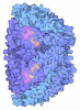
- Assembly
| Deposited unit | 
| ||||||||
|---|---|---|---|---|---|---|---|---|---|
| 1 |
| ||||||||
| Unit cell |
|
-Components
-Methyl-coenzyme M reductase subunit ... , 2 types, 4 molecules ADBE
| #1: Protein | Mass: 61535.535 Da / Num. of mol.: 2 / Source method: isolated from a natural source Details: The alpha subunit contains six post-translational modifications: Methylhistidine 268 Methylarginine 282 Hydroxytryptophane 439 Thioglycine 457 Didehydroaspartate 462 Methylcysteine 464 Source: (natural) Candidatus Methanoperedens sp. BLZ2 (archaea)Cell line: / / Organ: / / Plasmid details: / / Variant: / / Strain: BLZ2 / Tissue: / References: UniProt: A0A6A2FLY3, coenzyme-B sulfoethylthiotransferase #2: Protein | Mass: 45366.742 Da / Num. of mol.: 2 / Source method: isolated from a natural source Source: (natural) Candidatus Methanoperedens sp. BLZ2 (archaea)Cell line: / / Organ: / / Plasmid details: / / Variant: / / Strain: BLZ2 / Tissue: / References: UniProt: A0A6A2G3Q8, coenzyme-B sulfoethylthiotransferase |
|---|
-Protein , 1 types, 2 molecules CF
| #3: Protein | Mass: 28197.822 Da / Num. of mol.: 2 / Source method: isolated from a natural source Details: The gamma subunit contains a methylhistidine at position 159. Source: (natural) Candidatus Methanoperedens sp. BLZ2 (archaea)Cell line: / / Organ: / / Plasmid details: / / Variant: / / Strain: BLZ2 / Tissue: / References: UniProt: A0A5E4HJB0, coenzyme-B sulfoethylthiotransferase |
|---|
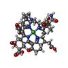
-Non-polymers , 8 types, 3454 molecules 


| #4: Chemical | ChemComp-NA /  Mass: 22.990 Da / Num. of mol.: 1 / Source method: obtained synthetically / Formula: Na Mass: 22.990 Da / Num. of mol.: 1 / Source method: obtained synthetically / Formula: Na | ||||||||||||
|---|---|---|---|---|---|---|---|---|---|---|---|---|---|
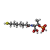
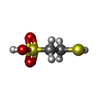
| #5: Chemical | ChemComp-EDO /  Mass: 62.068 Da / Num. of mol.: 21 / Source method: obtained synthetically / Formula: C2H6O2 Mass: 62.068 Da / Num. of mol.: 21 / Source method: obtained synthetically / Formula: C2H6O2#6: Chemical | ChemComp-NO3 /  Mass: 62.005 Da / Num. of mol.: 5 / Source method: obtained synthetically / Formula: NO3 Mass: 62.005 Da / Num. of mol.: 5 / Source method: obtained synthetically / Formula: NO3#7: Chemical |  Mass: 906.580 Da / Num. of mol.: 2 / Source method: obtained synthetically / Formula: C42H51N6NiO13 / Feature type: SUBJECT OF INVESTIGATION Mass: 906.580 Da / Num. of mol.: 2 / Source method: obtained synthetically / Formula: C42H51N6NiO13 / Feature type: SUBJECT OF INVESTIGATION#8: Chemical |  Mass: 343.334 Da / Num. of mol.: 2 / Source method: obtained synthetically / Formula: C11H22NO7PS / Feature type: SUBJECT OF INVESTIGATION Mass: 343.334 Da / Num. of mol.: 2 / Source method: obtained synthetically / Formula: C11H22NO7PS / Feature type: SUBJECT OF INVESTIGATION#9: Chemical |  Mass: 142.197 Da / Num. of mol.: 2 / Source method: isolated from a natural source / Formula: C2H6O3S2 / Feature type: SUBJECT OF INVESTIGATION Mass: 142.197 Da / Num. of mol.: 2 / Source method: isolated from a natural source / Formula: C2H6O3S2 / Feature type: SUBJECT OF INVESTIGATION#10: Chemical |  Mass: 39.098 Da / Num. of mol.: 2 / Source method: obtained synthetically / Formula: K Mass: 39.098 Da / Num. of mol.: 2 / Source method: obtained synthetically / Formula: K#11: Water | ChemComp-HOH / | Mass: 18.015 Da / Num. of mol.: 3419 / Source method: isolated from a natural source / Formula: H2O |
-Details
| Has ligand of interest | Y |
|---|---|
| Has protein modification | Y |
-Experimental details
-Experiment
| Experiment | Method: X-RAY DIFFRACTION / Number of used crystals: 1 |
|---|
- Sample preparation
| Crystal | Density Matthews: 2.2 Å3/Da / Density % sol: 44 % / Description: Thick, yellow, brick-shaped |
|---|---|
| Crystal grow | Temperature: 291.15 K / Method: vapor diffusion, sitting drop / pH: 8 Details: Crystallisation was carried out on a junior Clover plate using a solution containing 20% w/v polyethylene glycol 3,350, 50 mM Tris pH 8.0, and 200 mM potassium nitrate. The reservoir ...Details: Crystallisation was carried out on a junior Clover plate using a solution containing 20% w/v polyethylene glycol 3,350, 50 mM Tris pH 8.0, and 200 mM potassium nitrate. The reservoir contained 100 ul of crystallisation solution. 5 ul protein at 2.19 mg/ml in 25 mM Tris/HCl pH 8.0, 100 mM NaCl, 10% v/v glycerol and 2 mM dithiothreitol were mixed with 2 ul of the crystallisation solution. Crystals were soaked in a crystallisation solution supplemented with 20% ethylene glycol before freezing in liquid nitrogen. |
-Data collection
| Diffraction | Mean temperature: 100 K / Serial crystal experiment: N |
|---|---|
| Diffraction source | Source: SYNCHROTRON / Site: ESRF  / Beamline: BM07 / Wavelength: 0.9798 Å / Beamline: BM07 / Wavelength: 0.9798 Å |
| Detector | Type: DECTRIS PILATUS 6M / Detector: PIXEL / Date: Mar 30, 2023 |
| Radiation | Protocol: SINGLE WAVELENGTH / Monochromatic (M) / Laue (L): M / Scattering type: x-ray |
| Radiation wavelength | Wavelength: 0.9798 Å / Relative weight: 1 |
| Reflection | Resolution: 0.976→76.674 Å / Num. obs: 1130261 / % possible obs: 96.5 % / Redundancy: 7.8 % / CC1/2: 0.996 / Rmerge(I) obs: 0.119 / Rpim(I) all: 0.045 / Rrim(I) all: 0.128 / Net I/σ(I): 9.4 |
| Reflection shell | Resolution: 0.976→1.04 Å / Redundancy: 7.1 % / Rmerge(I) obs: 1.168 / Mean I/σ(I) obs: 1.7 / Num. unique obs: 56513 / CC1/2: 0.667 / Rpim(I) all: 0.468 / Rrim(I) all: 1.26 / % possible all: 70.9 |
- Processing
| Software |
| |||||||||||||||||||||||||||||||||||||||||||||||||||||||||||||||||||||||||||||||||||||||||||||||||||||||||||||||||||||||||||||||||||||||||||||||||||||||||||||||||||||||||||||||||||||||||||||||||||||||||||||||||||||||||
|---|---|---|---|---|---|---|---|---|---|---|---|---|---|---|---|---|---|---|---|---|---|---|---|---|---|---|---|---|---|---|---|---|---|---|---|---|---|---|---|---|---|---|---|---|---|---|---|---|---|---|---|---|---|---|---|---|---|---|---|---|---|---|---|---|---|---|---|---|---|---|---|---|---|---|---|---|---|---|---|---|---|---|---|---|---|---|---|---|---|---|---|---|---|---|---|---|---|---|---|---|---|---|---|---|---|---|---|---|---|---|---|---|---|---|---|---|---|---|---|---|---|---|---|---|---|---|---|---|---|---|---|---|---|---|---|---|---|---|---|---|---|---|---|---|---|---|---|---|---|---|---|---|---|---|---|---|---|---|---|---|---|---|---|---|---|---|---|---|---|---|---|---|---|---|---|---|---|---|---|---|---|---|---|---|---|---|---|---|---|---|---|---|---|---|---|---|---|---|---|---|---|---|---|---|---|---|---|---|---|---|---|---|---|---|---|---|---|---|
| Refinement | Method to determine structure: MOLECULAR REPLACEMENT / Resolution: 0.98→41.78 Å / SU ML: 0.06 / Cross valid method: FREE R-VALUE / σ(F): 1.34 / Phase error: 10.72 / Stereochemistry target values: ML Details: The model was built and corrected with COOT (Version 0.8.9.2)(52) and refined with PHENIX.refine. All atoms were considered anisotropic, with hydrogens added in riding mode. The model was ...Details: The model was built and corrected with COOT (Version 0.8.9.2)(52) and refined with PHENIX.refine. All atoms were considered anisotropic, with hydrogens added in riding mode. The model was validated by the MolProbity server (http://molprobity.biochem.duke.edu).
| |||||||||||||||||||||||||||||||||||||||||||||||||||||||||||||||||||||||||||||||||||||||||||||||||||||||||||||||||||||||||||||||||||||||||||||||||||||||||||||||||||||||||||||||||||||||||||||||||||||||||||||||||||||||||
| Solvent computation | Shrinkage radii: 0.9 Å / VDW probe radii: 1.1 Å / Solvent model: FLAT BULK SOLVENT MODEL | |||||||||||||||||||||||||||||||||||||||||||||||||||||||||||||||||||||||||||||||||||||||||||||||||||||||||||||||||||||||||||||||||||||||||||||||||||||||||||||||||||||||||||||||||||||||||||||||||||||||||||||||||||||||||
| Displacement parameters | Biso mean: 10.72 Å2 | |||||||||||||||||||||||||||||||||||||||||||||||||||||||||||||||||||||||||||||||||||||||||||||||||||||||||||||||||||||||||||||||||||||||||||||||||||||||||||||||||||||||||||||||||||||||||||||||||||||||||||||||||||||||||
| Refinement step | Cycle: LAST / Resolution: 0.98→41.78 Å
| |||||||||||||||||||||||||||||||||||||||||||||||||||||||||||||||||||||||||||||||||||||||||||||||||||||||||||||||||||||||||||||||||||||||||||||||||||||||||||||||||||||||||||||||||||||||||||||||||||||||||||||||||||||||||
| Refine LS restraints |
| |||||||||||||||||||||||||||||||||||||||||||||||||||||||||||||||||||||||||||||||||||||||||||||||||||||||||||||||||||||||||||||||||||||||||||||||||||||||||||||||||||||||||||||||||||||||||||||||||||||||||||||||||||||||||
| LS refinement shell |
|
