+ Open data
- Basic information
| Entry | Database: PDB / ID: 9qpw | |||||||||||||||||||||||||||||||||||||||||||||
|---|---|---|---|---|---|---|---|---|---|---|---|---|---|---|---|---|---|---|---|---|---|---|---|---|---|---|---|---|---|---|---|---|---|---|---|---|---|---|---|---|---|---|---|---|---|---|
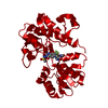
| Title | GluA4, resting state, structure of TMD/LBD | |||||||||||||||||||||||||||||||||||||||||||||
 Components Components | Isoform 2 of Glutamate receptor 4 | |||||||||||||||||||||||||||||||||||||||||||||
 Keywords Keywords | MEMBRANE PROTEIN / GluA4 / GRIA4 / TMD/LBD / Resting state / NBQX | |||||||||||||||||||||||||||||||||||||||||||||
| Function / homology |  Function and homology information Function and homology informationTrafficking of AMPA receptors / kainate selective glutamate receptor complex / regulation of synapse structure or activity / Synaptic adhesion-like molecules / Activation of AMPA receptors / AMPA glutamate receptor activity / Trafficking of GluR2-containing AMPA receptors / negative regulation of smooth muscle cell apoptotic process / AMPA glutamate receptor complex / ionotropic glutamate receptor complex ...Trafficking of AMPA receptors / kainate selective glutamate receptor complex / regulation of synapse structure or activity / Synaptic adhesion-like molecules / Activation of AMPA receptors / AMPA glutamate receptor activity / Trafficking of GluR2-containing AMPA receptors / negative regulation of smooth muscle cell apoptotic process / AMPA glutamate receptor complex / ionotropic glutamate receptor complex / Unblocking of NMDA receptors, glutamate binding and activation / positive regulation of synaptic transmission, glutamatergic / response to fungicide / glutamate-gated receptor activity / presynaptic active zone membrane / ligand-gated monoatomic ion channel activity involved in regulation of presynaptic membrane potential / synaptic transmission, glutamatergic / transmitter-gated monoatomic ion channel activity involved in regulation of postsynaptic membrane potential / postsynaptic density membrane / modulation of chemical synaptic transmission / terminal bouton / dendritic spine / chemical synaptic transmission / postsynaptic density / neuronal cell body / synapse / dendrite / glutamatergic synapse / identical protein binding / plasma membrane Similarity search - Function | |||||||||||||||||||||||||||||||||||||||||||||
| Biological species |  | |||||||||||||||||||||||||||||||||||||||||||||
| Method | ELECTRON MICROSCOPY / single particle reconstruction / cryo EM / Resolution: 3.8 Å | |||||||||||||||||||||||||||||||||||||||||||||
 Authors Authors | Vega-Gutierrez, C. / Herguedas, B. | |||||||||||||||||||||||||||||||||||||||||||||
| Funding support |  Spain, 1items Spain, 1items
| |||||||||||||||||||||||||||||||||||||||||||||
 Citation Citation |  Journal: Nat Struct Mol Biol / Year: 2025 Journal: Nat Struct Mol Biol / Year: 2025Title: GluA4 AMPA receptor gating mechanisms and modulation by auxiliary proteins. Authors: Carlos Vega-Gutiérrez / Javier Picañol-Párraga / Irene Sánchez-Valls / Victoria Del Pilar Ribón-Fuster / David Soto / Beatriz Herguedas /  Abstract: AMPA-type glutamate receptors, fundamental ion channels for fast excitatory neurotransmission and synaptic plasticity, contain a GluA tetrameric core surrounded by auxiliary proteins such as ...AMPA-type glutamate receptors, fundamental ion channels for fast excitatory neurotransmission and synaptic plasticity, contain a GluA tetrameric core surrounded by auxiliary proteins such as transmembrane AMPA receptor regulatory proteins (TARPs) or Cornichons. Their exact composition and stoichiometry govern functional properties, including kinetics, calcium permeability and trafficking. The GluA1-GluA3 subunits predominate in the adult forebrain and are well characterized. However, we lack structural information on full-length GluA4-containing AMPARs, a subtype that has specific roles in brain development and specific cell types in mammals. Here we present the cryo-electron microscopy structures of rat GluA4:TARP-γ2 trapped in active, resting and desensitized states, covering a full gating cycle. Additionally, we describe the structure of GluA4 alone, which displays a classical Y-shaped conformation. In resting conditions, GluA4:TARP-γ2 adopts two conformations, one resembling the desensitized states of other GluA subunits. Moreover, we identify a regulatory site for TARP-γ2 in the ligand-binding domain that modulates gating kinetics. Our findings uncover distinct features of GluA4, highlighting how subunit composition and auxiliary proteins shape receptor structure and dynamics, expanding glutamatergic signaling diversity. | |||||||||||||||||||||||||||||||||||||||||||||
| History |
|
- Structure visualization
| Structure viewer | Molecule:  Molmil Molmil Jmol/JSmol Jmol/JSmol |
|---|
- Downloads & links
- Download
| PDBx/mmCIF format |  9qpw.cif.gz 9qpw.cif.gz | 308.6 KB | Display |  PDBx/mmCIF format PDBx/mmCIF format |
|---|---|---|---|---|
| PDB format |  pdb9qpw.ent.gz pdb9qpw.ent.gz | 227.1 KB | Display |  PDB format PDB format |
| PDBx/mmJSON format |  9qpw.json.gz 9qpw.json.gz | Tree view |  PDBx/mmJSON format PDBx/mmJSON format | |
| Others |  Other downloads Other downloads |
-Validation report
| Arichive directory |  https://data.pdbj.org/pub/pdb/validation_reports/qp/9qpw https://data.pdbj.org/pub/pdb/validation_reports/qp/9qpw ftp://data.pdbj.org/pub/pdb/validation_reports/qp/9qpw ftp://data.pdbj.org/pub/pdb/validation_reports/qp/9qpw | HTTPS FTP |
|---|
-Related structure data
| Related structure data |  53285MC  9igzC  9qdnC  9rmsC  9rmwC  9rn4C  9rn7C  9rnhC M: map data used to model this data C: citing same article ( |
|---|---|
| Similar structure data | Similarity search - Function & homology  F&H Search F&H Search |
- Links
Links
- Assembly
| Deposited unit | 
|
|---|---|
| 1 |
|
- Components
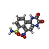
| #1: Protein | Mass: 98671.938 Da / Num. of mol.: 4 Source method: isolated from a genetically manipulated source Source: (gene. exp.)  Cell line (production host): Human embryonic kidney 293 cells Organ (production host): Kidney / Production host:  Homo sapiens (human) / References: UniProt: P19493 Homo sapiens (human) / References: UniProt: P19493#2: Chemical | ChemComp-E2Q / Has ligand of interest | Y | Has protein modification | Y | |
|---|
-Experimental details
-Experiment
| Experiment | Method: ELECTRON MICROSCOPY |
|---|---|
| EM experiment | Aggregation state: PARTICLE / 3D reconstruction method: single particle reconstruction |
- Sample preparation
| Component | Name: GluA4, resting state, structure of TMD/LBD / Type: COMPLEX / Entity ID: #1 / Source: RECOMBINANT | ||||||||||||||||||||
|---|---|---|---|---|---|---|---|---|---|---|---|---|---|---|---|---|---|---|---|---|---|
| Molecular weight | Value: 0.5 MDa / Experimental value: NO | ||||||||||||||||||||
| Source (natural) | Organism:  | ||||||||||||||||||||
| Source (recombinant) | Organism:  Homo sapiens (human) Homo sapiens (human) | ||||||||||||||||||||
| Buffer solution | pH: 8 | ||||||||||||||||||||
| Buffer component |
| ||||||||||||||||||||
| Specimen | Conc.: 4 mg/ml / Embedding applied: NO / Shadowing applied: NO / Staining applied: NO / Vitrification applied: YES Details: The sample was incubated with the AMPA receptor antagonist (NBQX) to capture the receptor in its resting state. | ||||||||||||||||||||
| Specimen support | Grid material: GOLD / Grid mesh size: 300 divisions/in. / Grid type: Quantifoil R1.2/1.3 | ||||||||||||||||||||
| Vitrification | Instrument: FEI VITROBOT MARK IV / Cryogen name: ETHANE / Humidity: 100 % / Chamber temperature: 277.15 K |
- Electron microscopy imaging
| Experimental equipment |  Model: Titan Krios / Image courtesy: FEI Company |
|---|---|
| Microscopy | Model: TFS KRIOS |
| Electron gun | Electron source:  FIELD EMISSION GUN / Accelerating voltage: 300 kV / Illumination mode: OTHER FIELD EMISSION GUN / Accelerating voltage: 300 kV / Illumination mode: OTHER |
| Electron lens | Mode: BRIGHT FIELD / Nominal defocus max: 2000 nm / Nominal defocus min: 800 nm |
| Specimen holder | Cryogen: NITROGEN / Specimen holder model: FEI TITAN KRIOS AUTOGRID HOLDER |
| Image recording | Electron dose: 50 e/Å2 / Film or detector model: GATAN K3 BIOCONTINUUM (6k x 4k) / Num. of real images: 25288 |
- Processing
| EM software |
| ||||||||||||||||||||||||||||
|---|---|---|---|---|---|---|---|---|---|---|---|---|---|---|---|---|---|---|---|---|---|---|---|---|---|---|---|---|---|
| CTF correction | Type: PHASE FLIPPING AND AMPLITUDE CORRECTION | ||||||||||||||||||||||||||||
| 3D reconstruction | Resolution: 3.8 Å / Resolution method: FSC 0.143 CUT-OFF / Num. of particles: 59714 / Symmetry type: POINT | ||||||||||||||||||||||||||||
| Atomic model building | Protocol: RIGID BODY FIT / Space: REAL Details: The TMD region of GluA4:G2restI and four GluA2 LBD monomers (PDB 3UA8) were rigid body fitted in Chimera. Manual model building, refinement and validation were performed using Refmac 5 ...Details: The TMD region of GluA4:G2restI and four GluA2 LBD monomers (PDB 3UA8) were rigid body fitted in Chimera. Manual model building, refinement and validation were performed using Refmac 5 Servalcat followed by model building in Coot and Real Space Refinement in Phenix. | ||||||||||||||||||||||||||||
| Atomic model building | 3D fitting-ID: 1 / Type: experimental model
| ||||||||||||||||||||||||||||
| Refine LS restraints |
|
 Movie
Movie Controller


 PDBj
PDBj


