 Movie
Movie Controller

[English] 日本語
 Yorodumi
- PDB-9qcg: Crystal structure of Methanopyrus kandleri malate dehydrogenase m... -
+ Open data

- Basic information
| Entry | Database: PDB / ID: 9qcg | ||||||
|---|---|---|---|---|---|---|---|
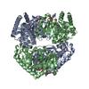
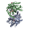
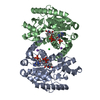
| Title | Crystal structure of Methanopyrus kandleri malate dehydrogenase mutant 4 at room temperature | ||||||
 Components Components | Malate dehydrogenase | ||||||
 Keywords Keywords | OXIDOREDUCTASE / malate dehydrogenase / Rossmann-like motif / NAD(P)H binding site | ||||||
| Function / homology |  Function and homology information Function and homology informationmalate dehydrogenase [NAD(P)+] / L-malate dehydrogenase (NADP+) activity / L-malate dehydrogenase (NAD+) activity / L-lactate dehydrogenase (NAD+) activity / lactate metabolic process / tricarboxylic acid cycle Similarity search - Function | ||||||
| Biological species |   Methanopyrus kandleri (archaea) Methanopyrus kandleri (archaea) | ||||||
| Method |  X-RAY DIFFRACTION / SYNCHROTRON / MOLECULAR REPLACEMENT / Resolution: 2.39 Å X-RAY DIFFRACTION / SYNCHROTRON / MOLECULAR REPLACEMENT / Resolution: 2.39 Å | ||||||
 Authors Authors | Coquille, S. / Madern, D. | ||||||
| Funding support |  France, 1items France, 1items
| ||||||
 Citation Citation | Journal: Mol.Biol.Evol. / Year: 2025 Title: Allostery and Evolution: A Molecular Journey Through the Structural and Dynamical Landscape of an Enzyme Super Family. Authors: Coquille, S. / Pereira, C.S. / Roche, J. / Santoni, G. / Engilberge, S. / Brochier-Armanet, C. / Girard, E. / Sterpone, F. / Madern, D. | ||||||
| History |
|
- Structure visualization
| Structure viewer | Molecule: MolmilJmol/JSmol |
|---|
- Downloads & links
-Download
| PDBx/mmCIF format | 9qcg.cif.gz | 289.4 KB | Display | PDBx/mmCIF format |
|---|---|---|---|---|
| PDB format | pdb9qcg.ent.gz | 191.5 KB | Display | PDB format |
| PDBx/mmJSON format | 9qcg.json.gz | Tree view | PDBx/mmJSON format | |
| Others |  Other downloads Other downloads |
-Validation report
| Arichive directory | https://data.pdbj.org/pub/pdb/validation_reports/qc/9qcgftp://data.pdbj.org/pub/pdb/validation_reports/qc/9qcg | HTTPS FTP |
|---|
-Related structure data
-Links
 PDBj
PDBj


- Assembly
| Deposited unit | 
| |||||||||||||||||||||||||||||||||||||||||||||||||||||||||||
|---|---|---|---|---|---|---|---|---|---|---|---|---|---|---|---|---|---|---|---|---|---|---|---|---|---|---|---|---|---|---|---|---|---|---|---|---|---|---|---|---|---|---|---|---|---|---|---|---|---|---|---|---|---|---|---|---|---|---|---|---|
| 1 | 
| |||||||||||||||||||||||||||||||||||||||||||||||||||||||||||
| Unit cell |
| |||||||||||||||||||||||||||||||||||||||||||||||||||||||||||
| Components on special symmetry positions |
| |||||||||||||||||||||||||||||||||||||||||||||||||||||||||||
| Noncrystallographic symmetry (NCS) | NCS domain:
NCS domain segments: Ens-ID: ens_1
NCS oper: (Code: givenMatrix: (-0.999992032861, 2.7752120961E-5, 0.00399167178221), (-3.9341511411E-5, -0.999995784516, -0.00290334341892), (0.00399157438144, -0.00290347732598, 0.999987818502)Vector: ...NCS oper: (Code: given Matrix: (-0.999992032861, 2.7752120961E-5, 0.00399167178221), Vector: |
-Components
| #1: Protein | Mass: 34652.887 Da / Num. of mol.: 2 / Mutation: D51H, R85Q, L146S, S228T, P232I Source method: isolated from a genetically manipulated source Source: (gene. exp.) Methanopyrus kandleri (archaea) / Gene: mdh, MK1069 / Production host:  #2: Chemical |   Mass: 745.421 Da / Num. of mol.: 2 / Source method: obtained synthetically / Formula: C21H30N7O17P3 Mass: 745.421 Da / Num. of mol.: 2 / Source method: obtained synthetically / Formula: C21H30N7O17P3#3: Chemical |   Mass: 35.453 Da / Num. of mol.: 3 / Source method: obtained synthetically / Formula: Cl Mass: 35.453 Da / Num. of mol.: 3 / Source method: obtained synthetically / Formula: Cl#4: Chemical |   Mass: 39.098 Da / Num. of mol.: 2 / Source method: obtained synthetically / Formula: K Mass: 39.098 Da / Num. of mol.: 2 / Source method: obtained synthetically / Formula: K#5: Water | ChemComp-HOH / |  Mass: 18.015 Da / Num. of mol.: 120 / Source method: isolated from a natural source / Formula: H2O Mass: 18.015 Da / Num. of mol.: 120 / Source method: isolated from a natural source / Formula: H2OHas ligand of interest | N | Has protein modification | N | |
|---|
-Experimental details
-Experiment
| Experiment | Method: X-RAY DIFFRACTION / Number of used crystals: 1 |
|---|
- Sample preparation
| Crystal | Density Matthews: 2.76 Å3/Da / Density % sol: 55.47 % |
|---|---|
| Crystal grow | Temperature: 293 K / Method: vapor diffusion, hanging drop Details: 0.2M potassium citrate tribasic monohydrate, 17% PEG3350 |
-Data collection
| Diffraction | Mean temperature: 293 K / Serial crystal experiment: N |
|---|---|
| Diffraction source | Source: SYNCHROTRON / Site: ESRF / Beamline: BM07 / Wavelength: 0.97951 Å |
| Detector | Type: DECTRIS PILATUS 6M / Detector: PIXEL / Date: Apr 20, 2023 |
| Radiation | Protocol: SINGLE WAVELENGTH / Monochromatic (M) / Laue (L): M / Scattering type: x-ray |
| Radiation wavelength | Wavelength: 0.97951 Å / Relative weight: 1 |
| Reflection | Resolution: 2.39→100 Å / Num. obs: 58746 / % possible obs: 99.9 % / Redundancy: 14 % / Biso Wilson estimate: 47.64 Å2 / CC1/2: 0.992 / Rmerge(I) obs: 0.648 / Net I/σ(I): 4.19 |
| Reflection shell | Resolution: 2.39→2.55 Å / Rmerge(I) obs: 4.549 / Mean I/σ(I) obs: 0.43 / Num. unique obs: 10401 / CC1/2: 0.323 |
- Processing
| Software |
| ||||||||||||||||||||||||||||||||||||||||||||||||||||||||||||||||||||||||||||||||||||
|---|---|---|---|---|---|---|---|---|---|---|---|---|---|---|---|---|---|---|---|---|---|---|---|---|---|---|---|---|---|---|---|---|---|---|---|---|---|---|---|---|---|---|---|---|---|---|---|---|---|---|---|---|---|---|---|---|---|---|---|---|---|---|---|---|---|---|---|---|---|---|---|---|---|---|---|---|---|---|---|---|---|---|---|---|---|
| Refinement | Method to determine structure: MOLECULAR REPLACEMENT / Resolution: 2.39→17.9 Å / SU ML: 0.4085 / Cross valid method: FREE R-VALUE / σ(F): 1.33 / Phase error: 36.5336 Stereochemistry target values: GeoStd + Monomer Library + CDL v1.2
| ||||||||||||||||||||||||||||||||||||||||||||||||||||||||||||||||||||||||||||||||||||
| Solvent computation | Shrinkage radii: 0.9 Å / VDW probe radii: 1.1 Å / Solvent model: FLAT BULK SOLVENT MODEL | ||||||||||||||||||||||||||||||||||||||||||||||||||||||||||||||||||||||||||||||||||||
| Displacement parameters | Biso mean: 55.9 Å2 | ||||||||||||||||||||||||||||||||||||||||||||||||||||||||||||||||||||||||||||||||||||
| Refinement step | Cycle: LAST / Resolution: 2.39→17.9 Å
| ||||||||||||||||||||||||||||||||||||||||||||||||||||||||||||||||||||||||||||||||||||
| Refine LS restraints |
| ||||||||||||||||||||||||||||||||||||||||||||||||||||||||||||||||||||||||||||||||||||
| Refine LS restraints NCS | Type: Torsion NCS / Rms dev position: 0.676903765272 Å | ||||||||||||||||||||||||||||||||||||||||||||||||||||||||||||||||||||||||||||||||||||
| LS refinement shell |
|
