Journal: Structure / Year: 2026 Title: Mechanism of SHP2 activation by bis-Tyr-phosphorylated Gab1. Authors: Lisa Machner / Alaa Shaikhqasem / Tobias Gruber / Farzad Hamdi / Constanze Breithaupt / Judith Kniest / Felix Wiebe / Marc Lewitzky / Christoph Parthier / Fotis L Kyrilis / Jochen Balbach / ...Authors: Lisa Machner / Alaa Shaikhqasem / Tobias Gruber / Farzad Hamdi / Constanze Breithaupt / Judith Kniest / Felix Wiebe / Marc Lewitzky / Christoph Parthier / Fotis L Kyrilis / Jochen Balbach / Panagiotis L Kastritis / Stephan M Feller / Milton T Stubbs / Abstract: The non-receptor tyrosine phosphatase SHP2 (SH2 domain-containing protein tyrosine phosphatase 2) (PTPN11) is a regulator of diverse cellular functions including mitogenic activation and cell ...The non-receptor tyrosine phosphatase SHP2 (SH2 domain-containing protein tyrosine phosphatase 2) (PTPN11) is a regulator of diverse cellular functions including mitogenic activation and cell migration. It comprises two tandem Src-homology 2 (SH2) domains followed by the catalytic domain and is autoinhibited by the N-terminal SH2 domain blocking access to the active site. Mutations influencing auto-inhibition have been implicated in cancer and other diseases, and allosteric inhibitors have been developed that stabilize the inactive state. Here, we show that the intrinsically disordered bis-phosphorylated SHP2-activating peptide pYpY-Gab1 binds to both SH2 domains, undergoing partial ordering in the process. In addition to eliciting changes in SH2 domain dynamics, the peptide reorganizes their relative orientations to generate a new SH2-SH2 interface. Our data suggest an active conformation for SHP2 that is also applicable to the hematopoietic cell-specific SHP1 (PTPN6), shedding light on the activation mechanism of both enzymes and paving the way for the development of novel compounds to modulate SHP2 activity.
In the structure databanks used in Yorodumi, some data are registered as the other names, "COVID-19 virus" and "2019-nCoV". Here are the details of the virus and the list of structure data.
Jan 31, 2019. EMDB accession codes are about to change! (news from PDBe EMDB page)
EMDB accession codes are about to change! (news from PDBe EMDB page)
The allocation of 4 digits for EMDB accession codes will soon come to an end. Whilst these codes will remain in use, new EMDB accession codes will include an additional digit and will expand incrementally as the available range of codes is exhausted. The current 4-digit format prefixed with “EMD-” (i.e. EMD-XXXX) will advance to a 5-digit format (i.e. EMD-XXXXX), and so on. It is currently estimated that the 4-digit codes will be depleted around Spring 2019, at which point the 5-digit format will come into force.
The EM Navigator/Yorodumi systems omit the EMD- prefix.
Related info.:Q: What is EMD? / ID/Accession-code notation in Yorodumi/EM Navigator
Yorodumi is a browser for structure data from EMDB, PDB, SASBDB, etc.
This page is also the successor to EM Navigator detail page, and also detail information page/front-end page for Omokage search.
The word "yorodu" (or yorozu) is an old Japanese word meaning "ten thousand". "mi" (miru) is to see.
Related info.:EMDB / PDB / SASBDB / Comparison of 3 databanks / Yorodumi Search / Aug 31, 2016. New EM Navigator & Yorodumi / Yorodumi Papers / Jmol/JSmol / Function and homology information / Changes in new EM Navigator and Yorodumi
 Movie
Movie Controller

 Yorodumi
 Open data
 Basic information
 Components
 Keywords
 Function and homology information
 Homo sapiens (human)
 X-RAY DIFFRACTION /
  Authors
 Germany, 1items
  Citation
 Structure visualization
 Downloads & links
 Other downloads
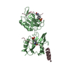
 PDBj
PDBj


 Assembly

 Mass: 18.015 Da / Num. of mol.: 43 / Source method: isolated from a natural source / Formula: H2O
 Sample preparation
 Processing